Recent developments in Microfluidics and biochemical techniques such as genetic identification and antibody capture have provided easier and more sensitive platforms for detecting and diagnosing diseases as well as providing new fundamental insight into disease progression. Recent years, Microfluidic sensors have attracted a wide attention because of the increased demands from the automation and control in microsystems. Various types of Microfluidic sensors have been developed for the Microfluidic systems or applications based on the research contributions in the last decade.
Optical-based Microfluidics
Due to their safety, rapidity, high sensitivity, and multipurpose capability, optical biosensors are very promising in biomarker detection methods. Optical properties including plasmonic, local surface plasmon resonance (SPR), localized SPR (LSPR) and surface enhanced Raman scattering (SERS) are usually used. Several classifications of optical detection techniques can be done based on the optical method, the imaging systems or the presence or not of lenses. Table 1 shows the most diffused optical techniques.
Table 1 The most diffused optical techniques
| Technique | Principle | Properties |
| Colorimetric Microfluidics | Measure of the attenuation of incident radiation in function of the wavelength |
|
| Fluorescence | Measure of emission light from a fluorophore |
|
| LIF/LEDIF | Excitation is caused by a laser or LED source |
|
| Chemiluminescence | Measure of energy release in a chemical reaction |
|
| SPR | Measure of refractive index change of a sample in contact with a metal film |
|
| Interferometers-based techniques | Measure of the phase shift caused by the analyte binding |
|
| SERS | Measure of plasmonic effect of metal substrates or nanoparticles |
|
| Lens-Less Imaging | Capture several such holograms by shifting the illumination source or the sample. |
|
Centrifugal Force-based Sensing
Centrifugal Microfluidics has evolved into a mature technology. The fields of application are widespread, including immunodiagnostics, protein analysis, and molecular diagnostics. Centrifugal Microfluidic technologies use the inertial pseudo forces experienced in a rotating reference frame to transport and manipulate fluids, overwhelmingly liquids, through networks of microchannels and chambers on substrates which are often in the format of a disk.
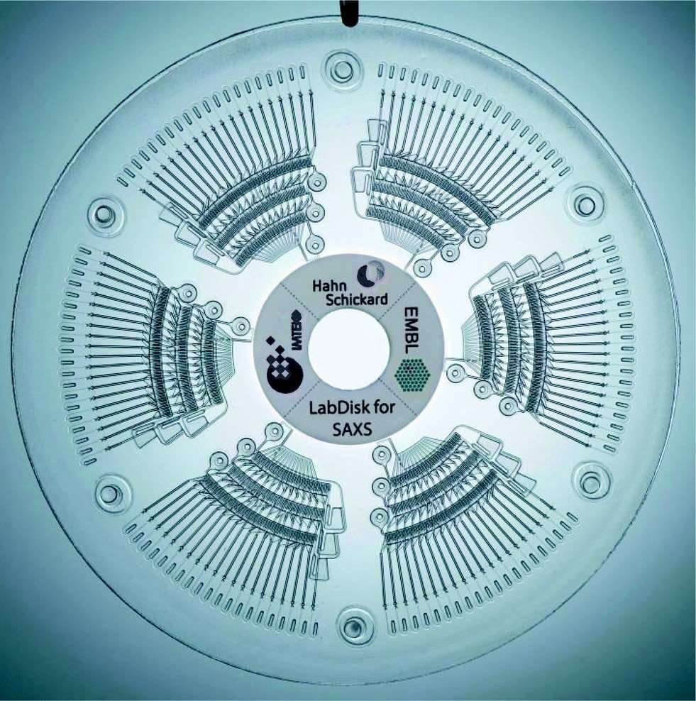 Fig.1 Schematic diagram of centrifugal micro-fluidic biochip.1
Electrical Sensing
To enable high-throughput cell analysis and non-invasive cell assays, electric systems have been incorporated into Microfluidic devices. Electric and electrochemical approaches are widely used for cell lysis and polymerase chain reaction (PCR)-based assays, including electrophoresis, dielectrophoresis (DEP), and electrowetting-on-dielectric (EWOD). For instance, by integrating the method of electrical cell-substrate impedance sensing with the Boyden chamber design, the state-of-the-art techniques provide kinetic information about tumor cell migration and invasion processes in 3D extracellular matrices.
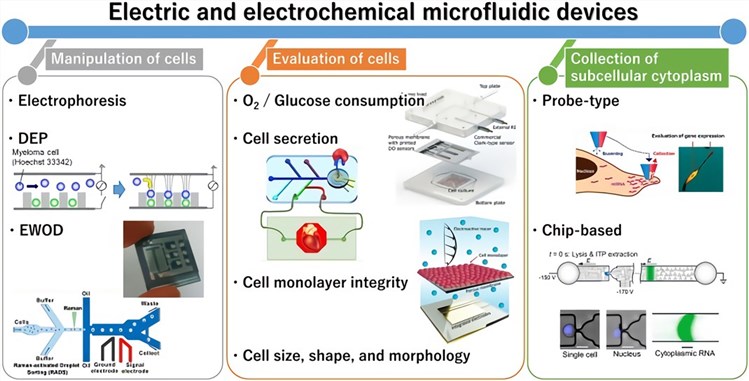 Fig.2 General outline of electric and electrochemical Microfluidic devices for cell analysis. (Hiramoto, 2019)
Creative Biolabs offers comprehensive Microfluidic chips-based services to support in vitro diseases research. For more information, please directly contact us and consult our technical supports online.
References
- Schwemmer, Frank, et al. "LabDisk for SAXS: a centrifugal microfluidic sample preparation platform for small-angle X-ray scattering." Lab on a Chip 16.7 (2016): 1161-1170.
- Hiramoto, K.; et al. Electric and electrochemical Microfluidic devices for cell analysis. Frontiers in Chemistry. 2019, 7: 396.
For Research Use Only.