From the read-out point of view, except for those tests that can be read by naked eye, most of the described platforms and technologies rely on optical or electrical detectors. Smartphones have evolved into powerful portable gadgets that integrate high processing power, together with physical sensors and connectivity. The image sensors within the smartphone’s camera module are sensitive enough for many diagnostically relevant applications. Portable imaging devices have the potential to improve screening and detection of disease in primary health care settings in both low- and high-resource countries. Point-of-care (POC) imaging is an alternative approach to laboratory-based analyses that provide diagnostic information in an outpatient setting, thereby reducing the time and infrastructure necessary for clinical decision-making. POC tests to meet specific ASSURED criteria: Affordable, Sensitive, Specific, User-friendly, Rapid and robust and Equipment-free.
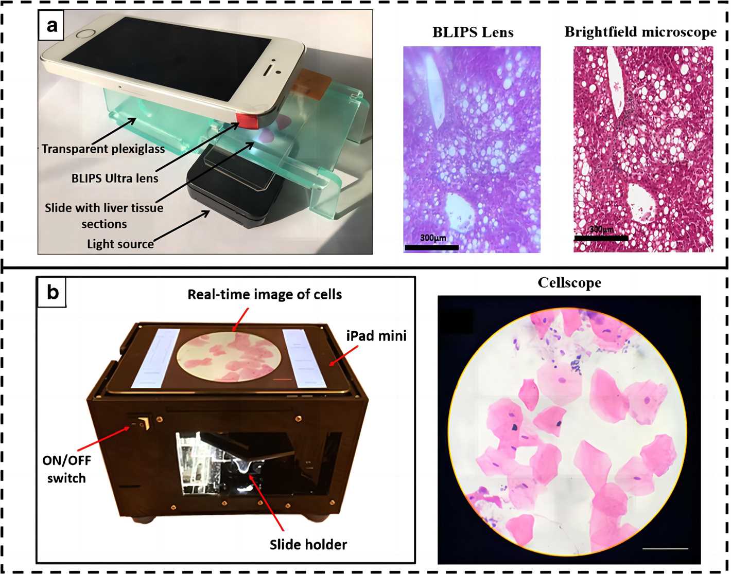 Fig.1 Smartphone-based imaging system.1, 4
Brightfield Microscopy
Smartphone cameras use low-cost complementary metal-oxide-semiconductor (CMOS) technology, which are able to detect red, green and blue (RGB) light and therefore ideal for optical quantification in the visible wavelength range. An initial application of the integrated CMOS sensors in smartphones was to use them as external cameras for capturing images from the microscope ocular using 3D-printed adapters. Furthermore, multiple studies have reported the design and development of attachments that are designed to comprise simplified optics that allow to perform microscopy observations directly. Perhaps the most unassuming yet most helpful of these applications is the smartphone brightfield microscope, which can be formed in a simple configuration by adding an external lens in front of the smartphone camera.
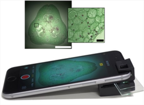 Fig.2 A smartphone accessory designed for brightfield microscopy.2, 4
Fluorescence Microscopy
Fluorescent microscopy is essential for modern biomedical diagnostics. To this application, smartphones have proven themselves to be more than capable of delivering diagnostically relevant results when equipped with the proper attachments. A typical smartphone fluorescence microscope consists of an excitation light source and an emission filter, in addition to the above-mentioned external lenses with the bright-field modality. How the samples are illuminated is the key consideration when designing a smartphone-based fluorescence microscope. For samples such as cuvettes or tubes, an orthogonal illumination to the detection path provides a simple means of high signal-to-noise ratio (SNR) fluorescence detection. For planar sample slides, waveguide coupling from the side of the glass substrate or from the end of a glass capillary tube has been proven to be an efficient background rejection method for sensitive fluorescence imaging. An alternative approach is the tilted illumination of the sample slides to achieve high SNR andtherefore improved detection sensitivity.
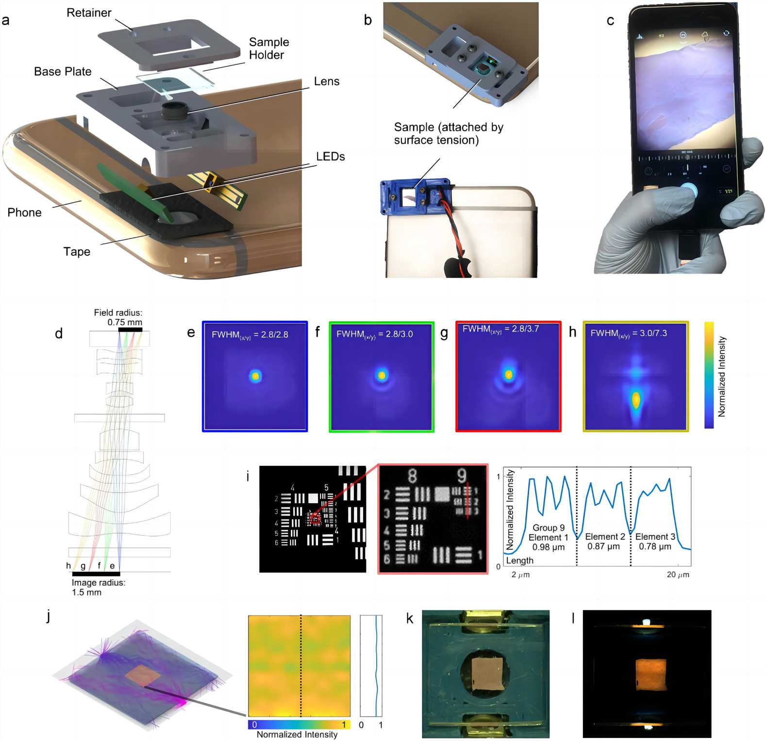 Fig.3 Compact smartphone microscope: practical implementation of microscopy with ultraviolet surface excitation.3, 4
Application of Microscopy in Smartphone-based POC
The detection of anthrax was enabled via a smartphone-based brightfield microscope that was used to monitor the growth of Bacillus anthracis spores on a Microfluidic incubation chip. This system was capable of detecting from 50 to 5000 spores in a period of 3–5 h. A compact smartphone-based microscope with the Newton Nm1 microscope, showed a performance comparable to a conventional microscope to detect Schistosoma mansoni and S. haematobium eggs in stool and stained urine samples. Similarly, a reversed-lens scope described above and a smartphonemounted microscope have been demonstrated for the detection of helminth eggs in stool and urine samples. Moreover, malaria parasites, sickled blood cells and tuberculosis (TB) bacilli have been captured with a high resolution with smartphone microscopes that include brightfield and fluorescence in blood and sputum samples.
References
- Banik, Soumyabrata, et al. "Recent trends in smartphone-based detection for biomedical applications: a review." Analytical and Bioanalytical Chemistry 413 (2021): 2389-2406.
- Hernández‐Neuta, Iván, et al. "Smartphone‐based clinical diagnostics: towards democratization of evidence‐based health care." Journal of internal medicine 285.1 (2019): 19-39.
- Liu, Yehe, et al. "Pocket MUSE: an affordable, versatile and high-performance fluorescence microscope using a smartphone." Communications biology 4.1 (2021): 334.
- Distributed under Open Access license CC BY 4.0, without modification.
For Research Use Only.