
The transition of exosome research from discovery to application requires rigorous validation of tissue restoration and functional integrity. Scalp health faces challenges from environmental pollutants, chemical treatments, and chronic micro-inflammation, making standardized, physiologically relevant data a critical component of any technical development dossier. Providing empirical evidence of a candidate's ability to assist in tissue recovery and the restoration of the innate defensive barrier is an essential step in the development of functional exosome preparations.
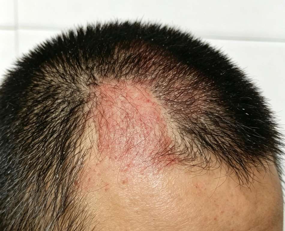
Creative Biolabs provides high-precision analytical solutions for biotechnology firms and cosmetic innovators. By utilizing a dual-model system, which combines high-resolution two-dimensional (2D) human keratinocyte assays with three-dimensional (3D) reconstructed human epidermal equivalents, we generate quantitative data to assess reparative claims. This service evaluates how exosome preparations may facilitate the transition from an injured state to a restored, homeostatic environment. By quantifying specific markers of repair, structural integrity, and innate immunity, we help bridge the gap between laboratory isolation and practical application.
Consult With Our Doctoral Team to Structure Your Scalp Repair Study
Our testing suite is built upon established biological pillars of dermatological repair, offering quantitative readouts for the recovery process.
When the scalp epidermal barrier is breached, the migration and proliferation of keratinocytes are primary requirements for closure. Exosomes carry microRNAs and signaling proteins that can activate intracellular pathways responsible for this movement. Our migration assays measure how exosome candidates influence the physical closure of an induced gap in a cellular monolayer.
Persistent inflammation is a significant obstacle to effective scalp repair. High levels of pro-inflammatory cytokines, such as Interleukin-6 (IL-6) and Tumor Necrosis Factor-alpha (TNF-α), are often observed in damaged tissue. We evaluate the potential of exosomes to assist in the down-regulation of these mediators to facilitate a regenerative state.
Authentic repair involves rebuilding the scalp's molecular shield. This involves the regulation of junctional proteins (Claudin-1, Zonula Occludens-1) and structural proteins (Keratin-1, Keratin-10). Our platform measures the density and distribution of these markers to verify structural recovery.
A functional repair process must restore the scalp's innate immune shield. Antimicrobial peptides (AMPs), such as Human Beta-Defensin 2 (hBD-2) and Cathelicidin (LL-37), are essential molecules produced by keratinocytes to protect the compromised barrier during recovery.
Request Detailed Scientific Protocols from Our Team
Our repair service is distinguished by its focus on dynamic injury modeling, providing a level of detail that generic screening protocols cannot achieve.

We offer specific injury induction protocols (UV-B irradiation or chemical stress) to measure active reparative responses rather than steady-state health.

By including the detection of antimicrobial peptides, we offer a more complete assessment of the functional repair of the scalp, moving beyond simple wound closure to evaluate the restoration of protective biological systems.

High-sensitivity ELISA provides a microscopic view of the inflammatory cascade to verify anti-inflammatory potency.

3D reconstructed human epidermal models provide a stratified architecture that resembles the human scalp, offering predictive data for in-vivo performance.
Connect With Our Project Consultant to Discuss Your Repair Study
Our laboratory follows a detailed, standardized workflow governed by strict Standard Operating Procedures (SOPs) to ensure every study is reproducible and defensible.
| Physicochemical Verification | Details |
|---|---|
| Physicochemical Verification |
The integrity of the exosome sample is verified upon receipt. We accept preparations from Mesenchymal Stem Cells (MSCs), botanical sources, or synthetic delivery systems.
|
| Internalization and Cellular Interaction | We confirm vesicle uptake into human keratinocytes over a 24 to 48 hour window using confocal microscopy. |
| The Reparative Assessment |
This phase involves the induction of scalp-relevant damage followed by the monitoring of the subsequent recovery through an evaluation of cellular and molecular indicators.
|
| Comprehensive Deliverables | Clients receive a technical dossier including raw data sets, high-resolution microscopy, and statistical significance analysis (P-values), providing the evidence needed for R&D decision-making or investor presentations. |
Submit Your Testing Requirement List to Our Project Consultant
Creative Biolabs' testing is centralized within a unified platform designed to eliminate experimental variance and ensure data integrity.



Access Platform Details with Our Specialists
Creative Biolabs' exosome offerings extend beyond scalp repair. Investigators can access a complete portfolio of hair and scalp efficacy assays, quality benchmarks, and adjunct bioengineering techniques, all structured to support laboratory research objectives and biological performance evaluations.
 Exosome extraction, purification and quantification
Retrieval and enrichment of exosomal vesicles from cultured media, biological liquids, or stem cell systems using standardized methods to guarantee lot to lot consistency and specified purity grades.
Exosome identification and quality analysis
Determination of physical attributes (size distribution and particle concentration) via NTA, morphological inspection through TEM, combined with immuno labeling of canonical exosomal markers including CD63, CD81, and TSG101.
Exosome profiling and proteomic detection
Characterization of protein constituents and nucleic acid payloads (e.g., miRNA, mRNA) to reveal molecular compositions and prospective signaling routes relevant to hair follicle biology.
Exosome engineering (optional)
Surface modification or cargo packaging approaches designed to improve targeted delivery characteristics and functional outcomes in follicular microenvironments.
Scalable exosome production
Generation of exosomes from adipose‑derived or other MSC sources at volumes adjustable to meet diverse research scales.
Exosome Anti-Hair Loss Efficacy Test
Assessment of follicular regeneration markers, anagen phase duration, and dermal papilla cell responsiveness.
Exosome Scalp Nourishment Efficacy Test
Assessment of barrier integrity and moisture maintenance.
Exosome Scalp Oil-Control Efficacy Test
Quantitative analysis of exosome‑mediated modulation of sebaceous gland function and sebum secretion for scalp surface homeostasis.
These services can be selected individually or as bundled panels, establishing a complete technical foundation for scalp repair efficacy investigations.
Get a Tailored Proposal for Your Exosome Candidate
A: Nourishment testing focuses on maintaining health and moisture in an already healthy tissue environment. Repair testing utilizes an injury-recovery model, where we deliberately stress the tissue (UV or chemical) and measure the exosome's ability to actively reverse that damage and restore barrier function.
A: TEWL is the gold-standard metric for barrier integrity. A high TEWL indicates a compromised scalp barrier. A reduction in TEWL following exosome application is direct evidence that the candidate is rebuilding the physical shield of the scalp and preventing excessive water evaporation.
A: During the repair process, the scalp is vulnerable to colonization. AMPs like hBD-2 and LL-37 represent the first line of innate defense. Evaluating these markers provides data on whether the candidate helps restore the biological shield that protects recovering tissue.
A: Yes. While our standard repair panel includes IL-6 and Claudin-1, we can substitute or add markers such as specialized Matrix Metalloproteinases or specific keratin subtypes depending on your research goals.
A: We test purified exosomes from MSCs, botanical, or microbial sources. If your candidate is supplied within a final formulation, please consult us for specialized preparation protocols.
Request A Scientific Consultation to Design Your Exosome Efficacy Study