Melanoma Tissue Exosome Research and Application
Introduction Our Expertise Scientific Background Platform Application Areas Workflow Advantages Deliverables Customer Feedback FAQ
Introduction: Advancing Melanoma Research Through Exosomal Insights
-
Melanoma is a highly aggressive skin cancer that originates from melanocytes. Although early-stage melanoma can often be treated with surgical resection, advanced stages, particularly metastatic melanoma, remain difficult to manage and are associated with poor prognosis.
-
In recent years, increasing attention has been given to tumor-derived exosomes as key mediators of intercellular communication. These nanoscale extracellular vesicles (30–150 nm) carry bioactive molecules—including proteins, RNAs, and lipids—that reflect the molecular signature of the originating tumor tissue.
-
Melanoma tissue-derived exosomes, unlike those from blood or cell lines, preserve more authentic tumor-specific cues, enabling researchers to study tumor progression, immune evasion, and microenvironment remodeling with greater biological relevance.
Creative Biolabs provides services for research and applications related to melanoma tissue exosomes to help clients better understand the relationship between exosomes and melanoma.
Our Expertise: Comprehensive Melanoma Exosome Analysis Services
At Creative Biolabs, we offer specialized services focused on isolating, purifying, and characterizing melanoma tissue-derived exosomes. Our workflows are optimized to preserve vesicle integrity and molecular content from even challenging samples such as formalin-fixed tissues or small biopsies.
Our services include:
-
Customized exosome isolation from fresh-frozen, FFPE, or cultured melanoma tissues
-
NTA (Nanoparticle Tracking Analysis), Western blotting, TEM imaging for exosome validation
-
Exosomal RNA sequencing (including miRNA profiling)
-
Mass spectrometry-based proteomics
-
Lipidomics for membrane composition analysis
-
Functional assays (e.g., immune modulation, angiogenesis, or migration)
Our team collaborates closely with academic and biotech clients to provide end-to-end experimental support, from study design through to data interpretation.
Scientific Background: The Role of Melanoma Exosomes in Tumor Biology
a. Melanoma Exosomes in Tumor Initiation and Progression
Melanoma-derived exosomes carry molecular cargo that promotes tumor cell proliferation, angiogenesis, and extracellular matrix (ECM) remodeling. For example:
-
miR-222 is highly expressed in melanoma exosomes, suppressing tumor suppressor genes like p27^Kip1 and activating the PI3K/AKT pathway.
-
let-7 family miRNAs (e.g., let-7a and let-7b) regulate integrin expression, cyclins, and CDK proteins, directly influencing melanoma growth and invasiveness.
-
Exosomal metalloproteinases (e.g., MMP-2, MMP-9) degrade ECM components, facilitating local invasion.
b. Melanoma Exosomes Facilitate Metastasis
Tissue-derived exosomes from highly metastatic melanoma strains can transfer invasive characteristics to low-metastatic tumor cells. Mechanistically:
-
They drive epithelial-mesenchymal transition (EMT) through miRNAs like miR-23a, miR-200a, and let-7i.
-
They activate MAPK signaling molecules such as MAP3K4 and MAPK13.
-
In sentinel lymph nodes, melanoma exosomes induce stromal remodeling and upregulate angiogenic molecules like uPA and collagen 18, preparing a favorable niche for metastasis.
c. Immune Suppression Mediated by Exosomes
Melanoma tissue exosomes are potent immunomodulators:
-
They express FASL on their surface, binding to FAS on CD4+ and CD8+ T cells and inducing apoptosis.
-
They downregulate anti-apoptotic proteins (BCL-2, BCL-xL, MCL-1) via miRNAs, further promoting immune cell death.
-
Additionally, melanoma exosomes interfere with dendritic cell maturation and T-cell activation, thereby suppressing adaptive immune responses.
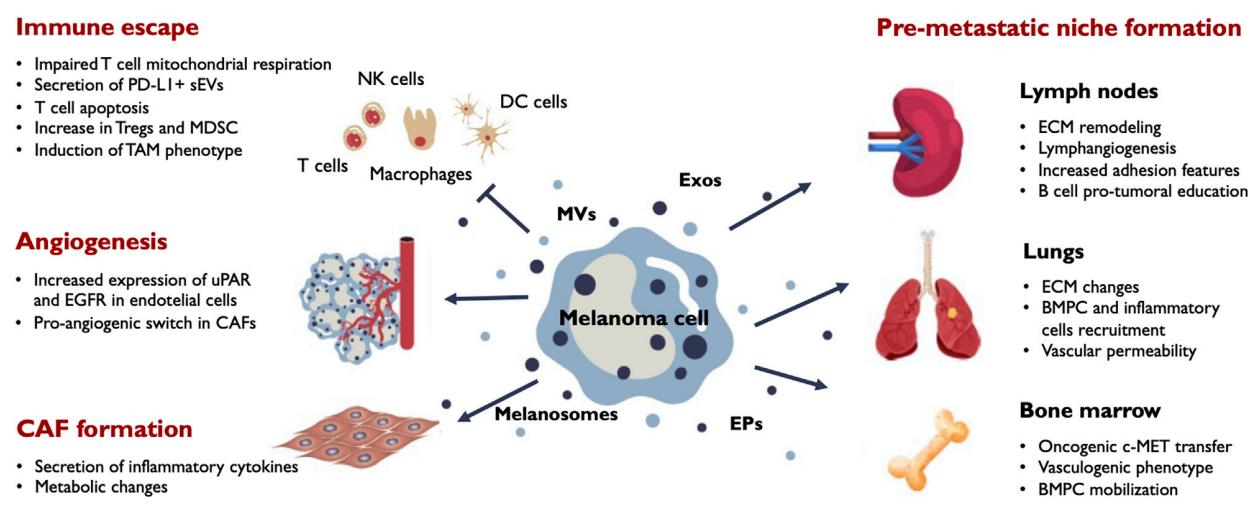 Fig.1 Main outcomes of EV release in melanoma progression.1
Our Technology Platform: Precision from Sample to Data
We employ a multi-tiered workflow optimized for high yield and purity:
-
Sample processing tailored to tissue type (fresh, FFPE, cryopreserved)
-
Exosome enrichment via ultracentrifugation, size exclusion chromatography, or immunoaffinity-based methods
-
Characterization using advanced tools (e.g., flow cytometry, cryo-TEM)
-
Multi-omics profiling to decode exosome content at the transcriptome, proteome, and lipidome levels
All procedures are performed in-house under rigorous QC standards, with results benchmarked against current literature and controls.
Application Areas: Advancing Melanoma Research
Our melanoma tissue exosome research platform supports diverse investigations, including:
-
Mechanistic studies on melanoma signaling and progression
-
Biomarker discovery for early detection and patient stratification
-
Preclinical validation of therapeutic strategies targeting exosome biogenesis or uptake
-
Exosome-based vaccine development for melanoma immunotherapy
-
Modeling tumor-host interactions in 3D co-culture or mouse models
Collaborators use our data to publish in high-impact journals, develop grant proposals, and validate novel therapeutic targets.
How We Work: From Tissue to Data — Your Research Pathway
-
Consultation & project design
-
Sample receipt and QC assessment
-
Exosome isolation and validation
-
Content analysis (RNA/protein/lipid)
-
Data delivery & interpretation
We maintain transparent communication throughout the process and offer flexible milestones to match your research timeline.
Advantages: Why Choose Creative Biolabs
-
Tissue expertise: Optimized for difficult tissue types such as melanoma biopsies and FFPE specimens
-
Integrated omics: Multi-dimensional analysis for robust biological insights
-
Data quality: Comprehensive QC at every stage
-
Customization: Fully adaptable services to your research goals
-
Experienced team: Scientists with expertise in exosome biology, melanoma research, and translational science
Deliverables: What You Receive
-
Detailed Reports: Comprehensive analysis of exosomal contents and their implications.
-
Raw Data Files: Access to all raw data for independent verification.
-
Consultation Services: Expert guidance on data interpretation and subsequent research steps.
Client Testimonials: Voices from the Research Community
"Collaborating with Creative Biolabs significantly advanced our understanding of melanoma exosomes. Their expertise and comprehensive analyses were instrumental in our research."
— Dr. JaXXX, Oncology Researcher
"The quality and depth of data provided by Creative Biolabs exceeded our expectations. Their team was responsive and highly knowledgeable."
— Dr. JoXXX, Immunology Department
Creative Biolabs is your reliable partner in exploring the complex biology of melanoma through tissue-based exosome studies. Our integrated approach empowers you to unravel the molecular mechanisms of melanoma with confidence. Contact us today to get started with a custom quote or scientific consultation.
Frequently Asked Questions
Q: What types of melanoma tissue samples are suitable for exosome analysis?
A: We accept fresh-frozen, FFPE, and cultured melanoma tissue samples. Each sample type undergoes optimized processing to ensure high-quality exosome isolation.
Q: How do you ensure the purity of isolated exosomes?
A: We implement a combination of ultracentrifugation, ultrafiltration, and size-exclusion chromatography, followed by rigorous quality assessments using NTA and electron microscopy.
Q: Can you assist with experimental design for exosome studies?
A: Absolutely. Our experts provide consultation services to help design experiments tailored to your research goals, including selecting appropriate controls and methodologies.
Q: What distinguishes melanoma-derived exosomes from those of non-malignant tissues?
A: Melanoma-derived exosomes often carry unique molecular signatures, including specific miRNAs and proteins that reflect the tumor's aggressive behavior, aiding in biomarker discovery and therapeutic targeting.
Q: How do the physical characteristics of exosomes influence their function?
A: Attributes like size, lipid composition, and surface markers affect exosome uptake by recipient cells and their role in intercellular communication, impacting tumor progression and immune modulation.
Reference
-
Benito-Martín, Alberto, Miriam Galvonas Jasiulionis, and Susana García-Silva. "Extracellular vesicles and melanoma: New perspectives on tumor microenvironment and metastasis." Frontiers in Cell and Developmental Biology 10 (2023): 1061982. Distributed under Open Access license CC BY 4.0. The image was modified by revising the title.
For Research Use Only. Cannot be used by patients.
Related Services: