Skin Organoid Introduction
Skin organoids are 3D skin-like structures generated by directing the differentiation of pluripotent stem cells (such as embryonic stem cells or induced pluripotent stem cells) or adult stem cells under specific conditions. Skin organoids typically contain epidermis and dermis layers alongside skin appendages including hair follicles, sweat glands and sebaceous glands. These structures have a high degree of similarity to the natural skin environment while partially reproducing skin functionalities. The formation of skin organoids occurs through self-organization which depends on inherent cellular features and external signaling signals like other organoids. Stem cells differentiate and proliferate throughout this process to create complex tissue structures.
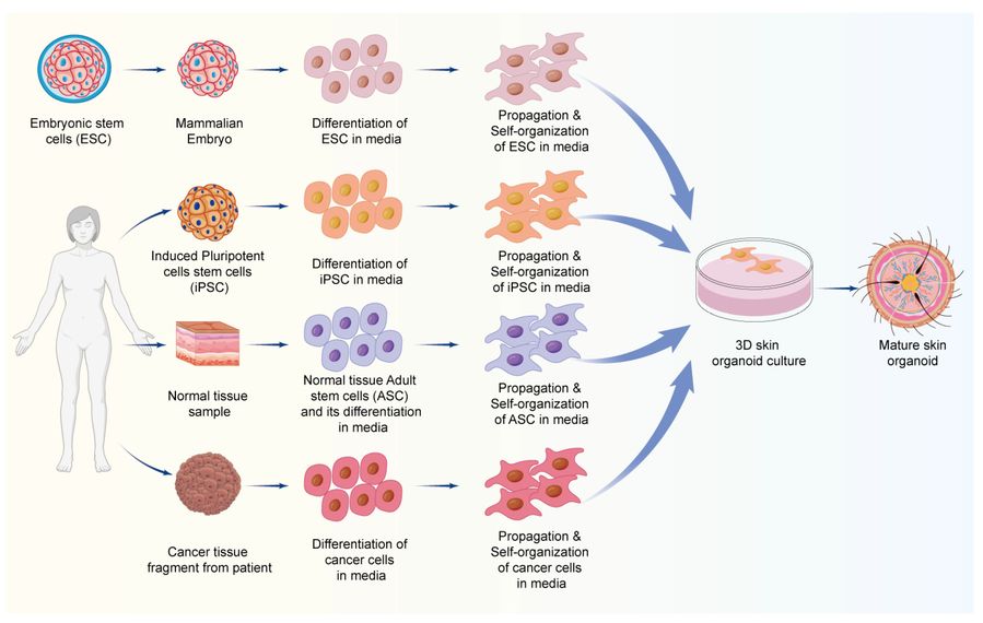 Figure 1 A schematic representation of the general process of skin organoid development.1
Comparative Analysis of Skin Organoids and Traditional Models
|
Feature
|
2D Cultures
|
Animal Models
|
Skin Organoids
|
|
Human Relevance
|
Low
|
Moderate
|
High
|
|
Structural Complexity
|
Minimal stratification
|
Species-specific
|
Native-like stratification
|
|
Appendage Formation
|
Absent
|
Present
|
Achievable (HF, SG, SW)
|
|
High-Throughput Screening
|
Excellent
|
Poor
|
Good
|
Characterization of Skin Organoids
-
Morphological Observation
Both optical microscopy and electron microscopy serve to evaluate skin organoids by examining their overall shape, size, and internal structural characteristics. Optical microscopy displays the stratified layers of the epidermis and skin appendages distribution while electron microscopy delivers detailed analysis of cellular morphology and intercellular connections.
-
Molecular Marker Detection
The examination of specific molecular markers enables identification of cell types and differentiation status in skin organoids. The expression of keratinocyte markers KRT10 and KRT14 helps identify epidermal cells while collagen I and collagen IV characterize dermal fibroblasts and specific markers like AGA and LCE detect skin appendage differentiation.
-
Functional Assays
Researchers can perform functional tests to determine the physiological activities present in skin organoids. TEWL measurements and skin barrier function assays help determine epidermal barrier function while dye uptake and secretion experiments assess sweat gland and sebaceous gland activity.
How to Design Skin Organoid Culture Medium?
Developing the ideal culture medium for skin organoids plays a critical role in directing differentiation processes while ensuring long-term viability and advancing maturation. The culture medium requires exact proportions of basal media along with essential nutrients and growth factors which must be supplemented by small molecules to match each developmental stage.
Basic Nutrients
Cells require amino acids, vitamins, and glucose in the culture medium to receive necessary metabolic components during both their growth and differentiation stages. Cells require these nutrients to generate energy and synthesize the essential components needed for cellular biosynthesis.
Growth Factors and Cytokines
Essential supplementation of skin development and homeostasis-related growth factors includes EGF, FGF, and PDGF. The behavior of skin cells including proliferation, differentiation and migration depends on regulatory factors.
Amino Acids
Amino acids are the basic building blocks of proteins and are essential for cell growth and proliferation. The culture medium should contain a variety of amino acids, such as glutamine, which is a key energy source for cells and plays a vital role in maintaining cell growth and metabolic activity.
Signaling Molecules
Constructing skin organoids from stem cells involves introducing signaling molecules that guide skin development through Wnt, Notch, and Hedgehog pathways. The development of epidermal structures and hair follicles relies on Wnt signaling as an essential pathway.
Skin Organoid Protocol: From Cells to Functional Models
A standardized protocol for skin organoid generation involves:
01 Cell Isolation
Primary keratinocytes and fibroblasts are harvested from skin biopsies or differentiated from hPSCs using defined growth factors (e.g., BMP4, FGF2).
02 Scaffold Selection
The cells are embedded in ECM components or decellularized dermal scaffolds to mimic the dermal-epidermal junction.
03 3D Culture
Organoids are cultured in serum-free media supplemented with EGF, FGF10, and insulin. Dynamic culture systems (e.g., bioreactors) enhance nutrient diffusion and maturation.
04 Maturation
Over 14-21 days, organoids develop multilayered epithelia with basal keratinocytes expressing K14 and suprabasal layers expressing K10.
Applications of Skin Organoids
Skin organoids have a wide range of potential applications in biomedical research and medicine:
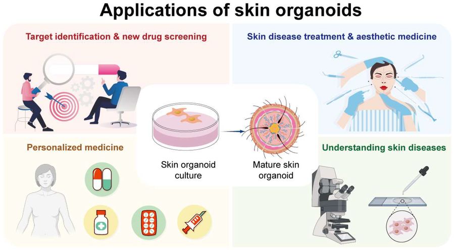 Figure 2 Applications of skin organoids: from culture to drug screening, aging research, and personalized medicine.1
Drug Discovery and Development
Skin organoids function as high-throughput screening systems to identify and develop new treatments for skin diseases and conditions. Testing therapeutic agents for efficacy and safety within skin organoids enables researchers to pinpoint potential drug candidates for subsequent evaluation in animal models and clinical trials. Researchers will save both time and money during drug development while enhancing the success probability of finding new drugs through this method.
Disease Modeling
Skin organoids can be used to model genetic skin disorders, autoimmune diseases of the skin, and a variety of skin cancers. Skin organoids with the molecular and cellular features of skin disorders are developed by researchers by patient-derived cells or by genetically modified cells. Disease mechanisms, diagnostic methods and therapeutic strategies can be studied and tested using personalized and relevant skin organoids.
Toxicity Testing
Skin organoids are useful in determining the safety of products, because they respond to toxins in a manner similar to humans. Skin organoids can be used to determine if the chemicals are safe for humans by applying the chemicals and environmental toxins onto the organoids and watching for toxic reactions that are harmful to humans. This would protect the consumers from the chemicals in the cosmetics and other products and also help protect the environment from the chemicals.
Frequently Asked Questions
Q: Are skin organoids vascularized or innervated?
A: Standard skin organoid protocols fail to naturally produce functional vascular networks and innervation. Developers in this domain continue to face this particular problem. Current research tackles the lack of vascular networks and innervation in skin organoids through methods that include co-culturing with endothelial cells and neural cells or utilizing microfluidic systems for vascularization and innervation induction.
Q: Can skin organoids replace animal testing for dermatological products?
A: Skin organoids represent a powerful human-based substitute for animal-based evaluations of skin irritation and sensitization as well as phototoxicity testing. As regulatory acceptance develops the sophisticated capabilities of skin organoids make them powerful candidates for replacing animal models or minimizing their use.
Q: What kind of skin diseases can be modeled using skin organoids?
A: Skin organoids enable the modeling of numerous skin diseases such as genetic disorders like epidermolysis bullosa and ichthyosis as well as inflammatory conditions including psoriasis and atopic dermatitis along with skin cancers like melanoma. Researchers gain significant advantages in modeling patient-specific diseases through the development of organoids from patient iPSCs.
Conclusion
Dermatological research enters a new era with skin organoids which surpass the constraints of conventional 2D cell cultures and animal models. Stem cell-derived self-organizing 3D structures replicate human skin's complex architecture and physiological functions with accurate epidermal and dermal layers and accessory structures such as hair follicles. The development of skin organoids creates new possibilities for tailored medical treatments and precise disease modeling of genetic and inflammatory skin conditions through improved drug discovery and toxicity testing while potentially reducing animal-based testing methods.
Transform Your Research Using Cardiac Organoid Models from Creative Biolabs
Skin organoids represent a highly promising research field with significant potential. Creative Biolabs is committed to advancing and applying skin organoid technologies, offering high-quality products and services to support researchers worldwide in their exploration of skin organoids.
Creative Biolabs offers a comprehensive range of skin organoid-related services. Leveraging advanced technologies and extensive expertise, the company provides skin organoid construction services using various cell sources, including adult stem cells. These services encompass custom culture medium development, optimized culture protocols, and high-quality skin organoid generation. Contact us today to learn more!
Reference
-
Wang X Y, Jia Q N, Li J, et al. Organoids as Tools for Investigating Skin Aging: Mechanisms, Applications, and Insights. Biomolecules, 2024, 14(11): 1436. https://www.mdpi.com/2218-273X/14/11/1436 (Distributed under Open Access license CC BY 4.0, without modification.)
Research Model
Related Sections: