Electrochemical biosensors overcome the limitations of miniaturization and cost presented in optical biosensors and opens the possibility to apply these devices to point-of-care (POC). Up to now, electrochemical biosensors have been used in the detection of various targets such as cancer biomarkers, chemotherapeutics or antibodies. Armed with avaliable practical strategies and successful completion of projects, Creative Biolabs is competent to develop electrochemical biosensors capable of rapidly and reliably detect the presence of interested targets for worldwide clients to support their research use.
Introduction
Biosensors arise as tools capable of producing continuous signals that are proportional to the number of binding molecules that react on the surface of a sensor. Generally, biosensors comprise three parts:
- The sensitive elements which are biologically derived material such as ssDNA.
- The transducer or detector element that transforms the detected signal into a readable, quantified output.
- The signal processor that displays the transformed signal in a user-friendly way.
Working Principle
The design of the electrochemical DNA biosensor is based on the hybridization dynamics of nucleic acid. When the target DNA contains a sequence that matches that of the immobilized oligonucleotide-probe DNA, a hybrid duplex DNA is formed at the electrode surface. Such a hybridization event is usually detected through the changed current signals of certain electroactive indicators, connected to the use of enzyme labels or redox labels, or from other hybridization-induced changes in electrochemical parameters.
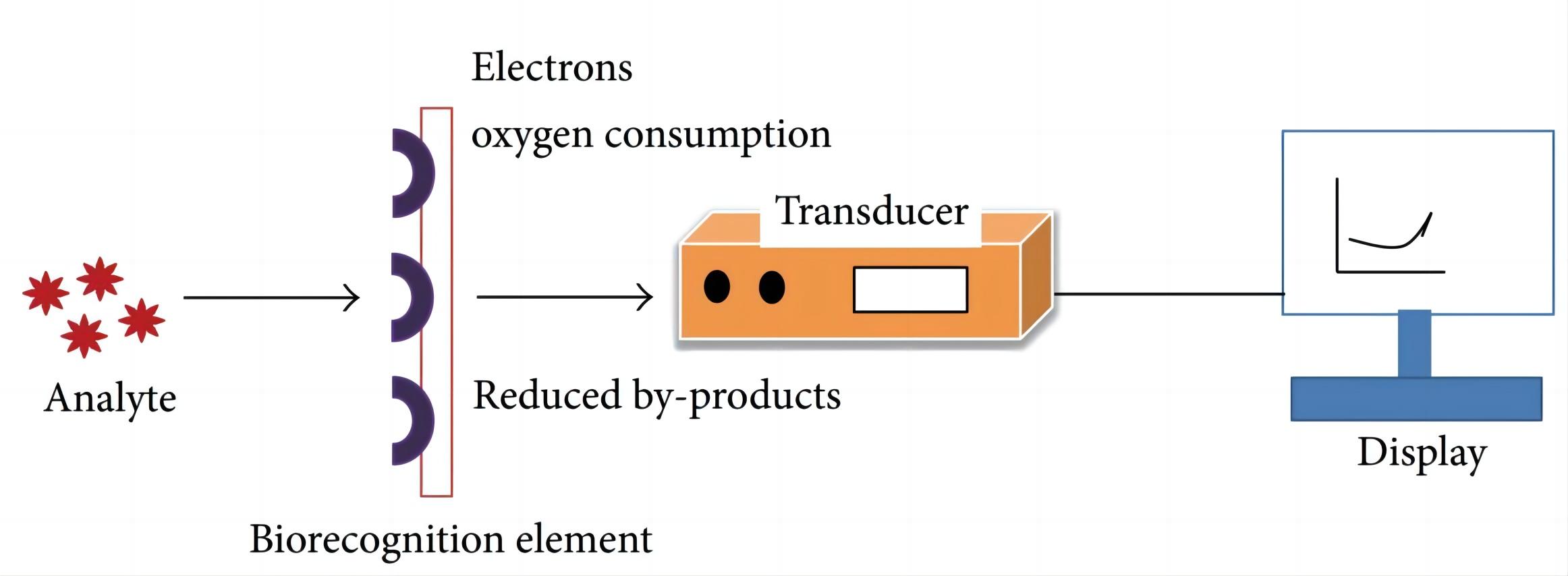 Fig.1 Basic principle of electrochemical biosensor.1
Important Considerations in Our Services
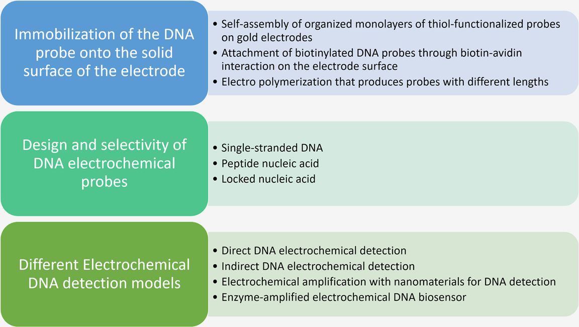
In order to guarantee good sensitivity of the biosensor, the stability of probes plays a very important role in the overall preparation of an electrochemical DNA biosensor. Besides, the design of the DNA electrochemical probe is the key factor in the fabrication of the electrochemical DNA biosensor. Creative Biolabs has accumulated a wealth of experience in years of project design work and optimized our biosensor development strategies. Many important factors, such as the ones listed below, should be taken into consideration.
Services
There are different methods to monitor DNA hybridization, including amperometry, voltammetry, potentiometry, and conductimetry. With the development of the integration of magnetic particles within biosensing devices, magnetic particles have been widely used in electrochemical biosensor. Pre-enrichment and purification are thus achieved simply by applying an external magnet. Creative Biolabs provides one-stop electrochemical biosensors for our honor clients. Some essential steps are listed as follows.
- Aptamer selection/modification
- Electrochemical biosensor design
- Fluorescence anisotropic analysis
- Electrode preparation and biosensor attachment
- Sensor behavior analysis
Creative Biolabs has addressed feasible methods to increase the sensitivity of electrochemical signals by implementing labeling techniques applicable to a variety of electroactive nanotracers. The electrochemical biosensors have been shown to be suitable for high-performance analysis in diverse field applications. If you are interested in our electrochemical biosensor development services, please feel free to contact us for more information.
Published Data
1. Development of Electrochemical Biosensor for p-Coumaric Acid Detection in Phytoproducts
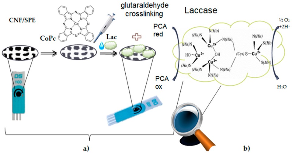 Fig.2 Preparation process of the CNF-CoPc-Lac/SPE Biosensor.2,4
This paper presented a novel enzymatic biosensor using a screen-printed electrode (SPE) modified with carbon nanofibers, cobalt phthalocyanine, and laccase (CNF-CoPc-Lac/SPE) for detecting p-coumaric acid (PCA) levels via cyclic voltammetry and square wave voltammetry. The sensor modification was achieved through casting and cross-linking techniques, utilizing glutaraldehyde as a reticulation agent. The biosensor demonstrated stable and sensitive responses to PCA redox processes. A calibration curve was developed for PCA concentrations ranging from 0.1–202.5 μM, with optimal performance observed in the 0.4–6.4 μM range. The sensor showed low limits of detection (LOD) and quantification (LOQ) values of 4.83 × 10⁻⁷ M and 1.61 × 10⁻⁶ M, respectively. PCA determination was successfully carried out in three complex phytoproducts, and voltammetric results were comparable to FTIR data. The CNF-CoPc-Lac/SPE biosensor could be employed for routine analysis of nutraceuticals, food products, and pharmaceuticals.
2. Paper-Based Electrochemical Biosensor for Detection of Procalcitonin
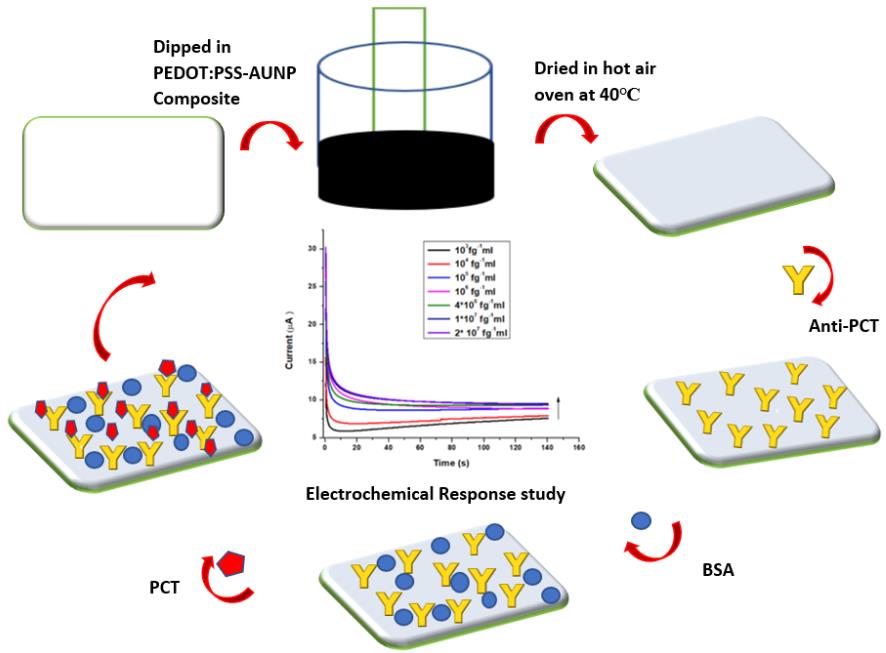 Fig.3 A schematic illustration depicting the fabrication process of a conducting paper-based PCT biosensor.3,4
In this research, an advanced cellulose fiber paper (CFP)-based biosensor was developed for the selective and sensitive detection of procalcitonin (PCT), a biomarker for bacterial infections. The sensor utilized a nanocomposite comprising poly(3,4-ethylene dioxythiophene) polystyrene sulfonate (PEDOT:PSS) and functionalized gold nanoparticles (PEDOT:PSS-AuNP@CFP). Characterization of the nanocomposite was done through Fourier transform infrared spectroscopy (FTIR), Scanning electron microscopy (SEM), and X-ray diffraction (XRD). This biosensor showed high sensitivity (1.34 μA (pg mL-1)−1) in the linear detection range of 1-20×104 pg mL−1 and a 24-day lifespan for PCT detection. Anti-PCT proteins were immobilized for PCT quantification. Electrochemical response studies demonstrated excellent reproducibility, stability, and sensitivity of this conductive paper bioelectrode in physiological ranges. The proposed bioelectrode offers a promising alternative for point-of-care PCT detection.
References
- Dhull, Vikas, et al. "Acetylcholinesterase biosensors for electrochemical detection of organophosphorus compounds: a review." Biochemistry research international 2013.1 (2013): 731501. Distributed under Open Access license CC BY 3.0, without modification.
- Bounegru, Alexandra Virginia, and Constantin Apetrei. "Development of a novel electrochemical biosensor based on carbon nanofibers–cobalt phthalocyanine–laccase for the detection of p-coumaric acid in phytoproducts." International Journal of Molecular Sciences 22.17 (2021): 9302.
- Gupta, Yachana, and Aditya Sharma Ghrera. "Development of conducting paper-based electrochemical biosensor for procalcitonin detection." ADMET and DMPK 11.2 (2023): 263-275.
- Distributed under Open Access license CC BY 4.0, without modification.
For Research Use Only.