Exosomes have the potential to contain individualized information related to disease state, therapeutic response, exposure to environmental cues, and a myriad of other health factors. With all these messages contained in a vesicular package that can be collected from bodily fluid samples, exosomes have generated considerable excitement as a pathway to enable personalized medicine. As a biotechnology company pursuing the frontier of science, Creative Biolabs has an advanced technology platform and we are capable of providing high-quality exosome detection services to global customers.
Introduction of Exosome in Liquid Biopsy
Exosomes are small (30-150nm) membrane-bound particles that originate from large multivesicular bodies (MVBs) and are released into the extracellular environment by fusion of MVBs with the plasma membrane, and they have been revealed as a promising biomarker in multiple diseases. Exosomes can also be released in large quantities in various biological fluids, such as plasma, urine, saliva, ascites, and bronchoalveolar lavage fluid. A number of cell types have been described as releasing exosomes, such as epithelial cells, hematopoietic cells, neuronal cells, fibroblasts, adipocytes, and tumor cells. Although the size of exosomes is similar between normal and malignant cells, the exosome protein concentration is higher in advanced-stage cancer, and the protein, mRNA, and miRNA profiles of exosomes also differ from those of exosomes cells of origin. These specific exosomes can alter the microenvironment through their protein and RNA cargo. Thus, in light of their crucial role in cell-cell communication and tumor-specific content, exosomes represent a promising biomarker for early tumor detection and monitoring and medication planning.
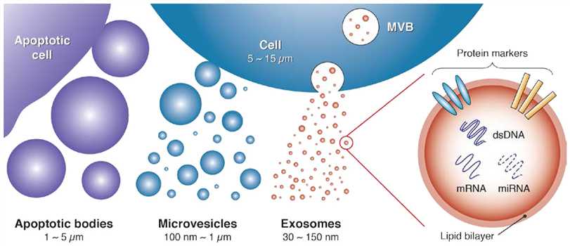 Fig.1 Schematic of exosomes, a subset of extracellular vesicles.1
Technology for Detecting Exosomes
The isolation of exosomes ideally not only yields large quantities of purified exosomes but also facilitates analysis, such as analysis of the proteins, DNA, and miRNA in exosomes.
- Isolation methods of exosome
Conventional methods of exosome isolation are based on a series of centrifugation steps to exclude cells and cell debris. In addition, exosomes can be isolated via ultrafiltration plus size exclusion chromatography, precipitation with polymers, and immunoaffinity purification using magnetic beads. Each method has advantages and drawbacks. For example, UC-SEC uses a solid matrix to isolate highly purified exosomes but it is difficult to remove contaminating proteins. Polymeric precipitation functions by capturing exosomes of a certain size (60-150nm) in 30 min and yields more exosomes than ultracentrifugation; however, it cannot avoid mixing of non-exosomal contents with a similar size, such as apoptotic debris or other types of microvesicles. Exosomes can also be isolated using an immunoaffinity method to selectively isolate classic CD9+, CD63+, or CD81+ exosomes, with the captured exosomes retaining bioactivity for downstream analysis. The immunoaffinity method can only be applied to a small-volume sample and only isolates exosomes with specific markers, which may limit the experimental findings.
- Analysis methods of exosomes
Exosomes are rich in RNA transcripts; thus, following isolation and validation via electron microscopy, the RNA contents of exosomes can be analyzed using qPCR and NGS. In addition to RNA, several types of protein have been reported to play important roles in cell-cell communication. Using proteomic techniques, such as mass spectrometry, researchers identified that a cell surface proteoglycan, glypican-1 (GPC1), could be applied as an exosome marker in breast cancer and pancreatic cancer patients.
Services at Creative Biolabs
Committed to the research of liquid biopsy over years, Creative Biolabs has step-by-step equipped our platform with advanced facilities, the latest technologies, and professional experts. With a comprehensive platform, we are confident in offering reliable exosome detection services to global researchers. In addition, our team of Ph.D. level scientists will help to deal with every problem during the projects to assure the quality of our services.
As an industry-leading biotechnology company, Creative Biolabs has strong foundations and mature technologies. We aspire to bring top-rated customer experiences to every client and we are doing so. If you are interested in exosome detection services or you have any other questions about our services, please don't hesitate to contact us for more information.
Published Data
1. Exosome Liquid Biopsy for Pre-Operative Detection of Lymph Node Metastasis in High-Risk T1 Colorectal Cancer
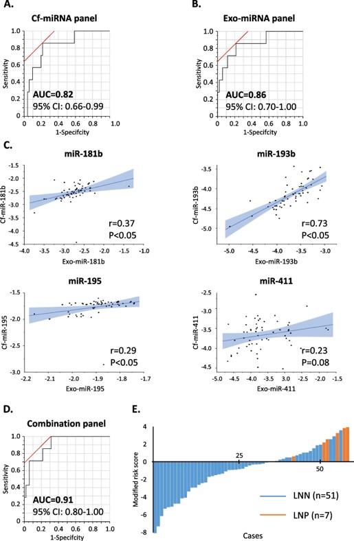 Fig.2 Training of exosomal miRNAs and cell-free miRNAs for identifying LNM.2,4
This study explored exosomal miRNAs as potential biomarkers for predicting lymph node metastasis (LNM) in submucosal colorectal cancer (T1 CRC). Researchers analyzed serum samples from 200 high-risk T1 CRC patients across two cohorts (training: 58, validation: 142). They extracted cell-free and exosomal RNAs and used qRT-PCR to examine miRNA panels. A combination of four miRNAs (miR-181b, miR-193b, miR-195, miR-411) demonstrated strong predictive ability for LNM, particularly in exosomal RNA. The resulting cell-free and exosomal miRNA signature was validated with an AUC of 0.84 (95% CI 0.70–0.98). A risk-stratification model incorporating pathological features reduced false positive rates for LNM by 76%, with no missed true LNM cases. This exosomal miRNA-based liquid biopsy could help identify T1 CRC patients at risk of LNM, potentially minimizing unnecessary treatments.
2. Exosomal Thomsen–Friedenreich (TF) Glycoantigen as a Novel Liquid Biopsy Biomarker for the Diagnosis of Lung and Breast Cancers
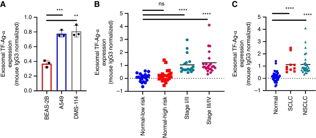 Fig.3 Evaluation of exosomal TF-Ag-α in lung cancer diagnosis.3,4
In this study, researchers identified a novel exosomal biomarker, α-linked Thomsen–Friedenreich glycoantigen (TF-Ag-α), and designed a surface plasmon resonance-based assay using a monoclonal antibody to detect its levels in the blood. They are the first to show that exosomes carry TF-Ag-α. The biomarker was detectable in serum samples as small as 10 μL from cancer patients, while levels were negligible in normal controls. In a cohort of 233 individuals (cancer patients and normal controls), exosomal TF-Ag-α accurately detected lung cancer (n = 60) with≥95% sensitivity and breast cancer (n = 95) with≥97% sensitivity. These results proved it a highly sensitive and specific cancer diagnostic biomarker.
References
- Contreras-Naranjo, Jose C., Hung-Jen Wu, and Victor M. Ugaz. "Microfluidics for exosome isolation and analysis: enabling liquid biopsy for personalized medicine." Lab on a Chip 17.21 (2017): 3558-3577. Distributed under Open Access license CC BY 3.0, without modification.
- Miyazaki, Katsuki, et al. "An exosome-based liquid biopsy signature for pre-operative identification of lymph node metastasis in patients with pathological high-risk T1 colorectal cancer." Molecular Cancer 22.1 (2023): 2.
- Hsu, Chang-Chieh, et al. "Exosomal Thomsen–Friedenreich Glycoantigen: A New Liquid Biopsy Biomarker for Lung and Breast Cancer Diagnoses." Cancer Research Communications 4.8 (2024): 1933-1945.
- Distributed under Open Access license CC BY 4.0, without modification.
For Research Use Only.