Creative Biolabs is a well-recognized expert in the field of antibody generation and production. Especially, we have launched a series of in vitro diagnostic (IVD) antibody development services for different infections and diseases. Scientific progress has resulted in the discovery of novel disease biomarkers to fulfill the need for a quicker, more specific and more accurate diagnosis. Particularly, we provide IVD antibody development services against glutathione-s-transferase.
Glutathione-s-Transferase
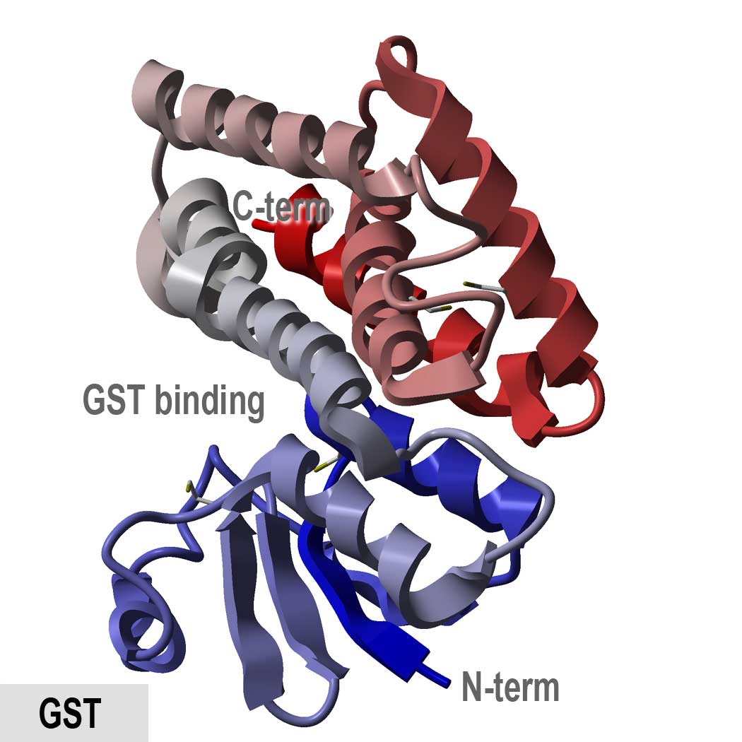
Glutathione-s-transferases (GSTs), previously being called ligandins, consist of a family of eukaryotic and prokaryotic phase II metabolic isozymes, which are widely known for their capability to catalyze the conjugation of the reduced form of glutathione (GSH) to xenobiotic substrates for the sake of detoxification. The GST family comprise three superfamilies: the mitochondrial proteins cytosolic proteins, and microsomal proteins—alternatively known as MAPEG. GSTs may comprise up to 10% of the cytosolic protein in several mammalian organs. As through a sulfhydryl group, GSTs can catalyze the conjugation of GSH to electrophilic centers on various substrates for the purpose of making the compounds more water-soluble. This process detoxifies endogenous compounds like peroxidised lipids and permits the breakdown of xenobiotics. GSTs might also bind toxins and act as transport proteins, which lead to the early term for GSTs, ligandin.
 Fig.1 Structure of glutathione s-transferase.Distributed under public domain, from Wiki,
without modification.
Diagnostic Utility of GSTs in Kidney Disease
The GST protein family is divided into three major subclasses α, π, and μ, while the α and π forms exist in human kidney with relatively high amounts in renal tubules. Urinary GSTs are well validated, histologically defined renal biomarkers whose cellular origins are the key to their diagnostic interpretation. Urinary GSTs are well validated and histologically defined renal biomarkers since their cellular origins are essential to interpret their diagnostic. The GST proteins are site-specific in the nephron: the α-GST isoform is primarily localized in the proximal tubule while the π-GST is in the distal tubule. α-GST and π-GST are preformed cytosolic enzymes and constitute 2% of soluble protein in the renal tubules. And these biomarkers are completely released into the urine during the renal injury, which enables them to be extremely early indicators of tubular damage. Therefore, the measurement of urinary α-GST and π-GST allow a topological diagnostic approach of renal injury due to the site-specific expression of these proteins in the renal tubules.
IVD Antibody Development Services Targeting GSTs
Antibodies are core elements for antibody-based immunoassays for detecting and quantifying antigens of interest in all kinds of samples such as the serum, urine, tissue preparations, and so on. IVD antibodies are extensively used for disease screening, prognosis, and therapeutic monitoring. With our versatile IVD platform, Creative Biolabs is proud to develop novel anti-GST antibody from scratch to commercial IVD kit (we can also start with provided antibody candidates). If you are interested in our services, please do not hesitate to contact us for more details.
For Research Use Only.