Creative Biolabs has all along focused on the development of in vitro diagnostic (IVD) antibodies for the diagnosis of a wide range of diseases. We’re capable of offering high-quality antibody development services against specific biomarkers of brain cancer to provide important information for its prognosis, diagnosis, and treatment. In particular, Huntingtin interacting protein 1 (HIP1) is a potential biomarker for brain cancer.
Introduction of HIP1
HIP1, also known as SHONgamma, is a 116-kD endocytic protein which belongs to an evolutionarily conserved family. It is required for the normal cell growth and when over-expressed, transforms into fibroblasts. HIP1 was originally characterized by a yeast two-hybrid system in 1997 and was a component of the clathrin-mediated endocytosis pathway due to its association with both the actin cytoskeleton and endocytosis mechanism. HIP1 and its only known mammalian homolog HIP1-related protein (HIP1r) are implicated in neurodegeneration based on the findings that HIP1 interacts with the Huntington protein ( mutated in Huntington's disease), and acts as a part of the endocytic machinery by binding to clathrin, actin, and AP2. Furthermore, many studies have suggested that HIP1 plays a role in tumorigenesis by the evidence of overexpression in various human malignancies and the transformation into fibroblasts.
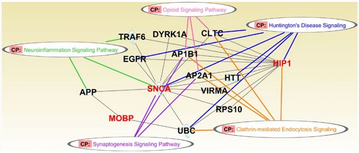 Fig.1 HIP1 in multiple system atrophy.1
HIP1 Marker for Brain Cancer
Brain tumors are the growth of malignant cells in tissues of the brain. There are more than 120 different classes of brain tumors, including the glioma (astrocytoma, glioblastoma multiforme (GBM)), pituitary tumor, and many others. The upregulation of HIP1 in glioblastoma and oligodendroglioma causes a prolonged half-life of growth factor receptors, such as epidermal growth factor receptor (EGFR) and platelet-derived growth factor β receptor (PDGF-βR). Moreover, EGFR and its downstream pathways are essential for the invasion and proliferation of tumor cells.
It has been reported by Bradley et al. (2007) that HIP1 is overexpressed in primary brain tumors. Tissue microarrays with 75 normal cortical brain tissue and 78 brain cancer tissue samples were stained for HIP1. The spots were scored as positive or negative (score <2) for HIP1 expression, with standards of 0 (no staining), 1+ (low staining), 2+ (intermediate staining), and 3+ (high staining). Fig.2A displays a GBM with 3+ positive HIP1 stain; a GBM with a 2+ positive HIP1 stain; a pilocytic astrocytoma with a 3+ positive HIP1 stain; and an oligodendroglioma with a 1+ negative stain. Overall, the frequency of HIP1 level was significantly higher in primary brain tumor tissues than in normal cortical brain tissues. Additional evidence indicated that common glial tumors tended to express HIP1 more frequently than less common brain tumors, such as medulloblastoma, ependymoma, pilocytic astrocytoma, and peripheral nerve sheath tumors.
IVD Antibody Development Services Targeting HIP1 Marker
HIP1 is overexpressed in a number of cancers, including brain, colon, and breast cancers. The detection of HIP1 can help the diagnosis and prognosis of these diseases. Therefore, we offer customized services for industries and research scientists that are interested in developing HIP1 antibodies for brain cancer diagnosis and prognosis. Moreover, these antibodies can be used for immunoassay development, followed by further validation for diagnostic uses. With our versatile IVD platform, Creative Biolabs is proud to develop novel HIP1-specific (paired) antibodies from scratch to commercial IVD kit (we can also start with provided antibody candidates).
If you’re interested in our services, please contact us for more information or directly send an inquiry.
Reference
- Bettencourt, Conceição, et al. "MOBP and HIP1 in multiple system atrophy: New α‐synuclein partners in glial cytoplasmic inclusions implicated in the disease pathogenesis." Neuropathology and applied neurobiology 47.5 (2021): 640-652. Distributed under Open Access license CC BY 4.0, without modification.
For Research Use Only.