Therapeutic Exosomes for Hepatocellular Carcinoma
Hepatocellular Carcinoma Exosome Delivery Advantages Research Our Services Available Products FAQs
Pathological Complexity and Current Barriers in Hepatic Oncology
Malignant epithelial tumors of the liver present a formidable challenge to global public health, currently ranking as the sixth most prevalent malignancy worldwide. Within primary liver cancers, hepatocellular carcinoma (HCC) constitutes approximately 80% of all diagnosed cases, reflecting a high incidence and a critical need for advanced therapeutic strategies. The etiology of this disease is heavily linked to chronic inflammatory stimuli and metabolic derangements, with major risk factors including persistent hepatitis B virus (HBV) and hepatitis C virus (HCV) infections, chronic exposure to aflatoxins, excessive alcohol consumption, obesity, tobacco smoking, and type 2 diabetes.
Despite iterative advancements in surgical resection techniques, local ablation, and systemic molecular targeted therapies, the clinical prognosis for individuals diagnosed with this malignancy remains poor. The five year overall survival rate continues to hover below 25%. This clinical failure is largely driven by three distinct pathological factors:
Asymptomatic Early Progression
The lack of specific early stage symptoms often lead to diagnosis at advanced stages, where curative surgical options are no longer applicable.
Intensified Chemoresistance
Malignant hepatocytes display a high tolerance to standard systemic chemotherapeutic agents, rendering conventional cytotoxic regimens ineffective.
Proangiogenic Tumor Microenvironment
The liver parenchyma under chronic disease states undergoes aggressive vascular remodeling, facilitating rapid intrahepatic metastasis and frequent recurrence post resection.
Consult With Our Scientists to Define Your Hepatic Target Parameters
Biophysical Suitability of Endogenous Bilayers for Targeted Cargo
The persistence of hepatic malignancies is partly maintained by an intricate network of intercellular communication within the tumor niche. In this microenvironment, endogenous nanovesicles, specifically exosomes, serve as vital transport units. These structures selectively package microRNAs (miRNAs) that act on various oncogenes and tumor suppressors, thereby driving malignant transformation, stromal remodeling, angiogenesis, and drug resistance. However, the exact biophysical mechanisms that allow these natural vesicles to coordinate tumor progression can be repurposed to design target specific drug delivery configurations.
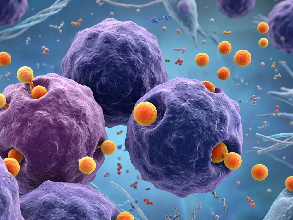
Utilizing lipid bilayers of endosomal origin as biological carriers offers distinct advantages over conventional synthetic nanostructures, such as synthetic liposomes or polymeric nanoparticles, particularly when targeting the hepatic filtration system:
Innate Vascular Stability
Derived from natural cellular membranes, these vesicles possess a specialized surface chemistry that allows them to circulate through the bloodstream without inducing premature macrophage clearance.
Low Baseline Immunogenicity
These carriers do not trigger hyper inflammatory responses or complement activation, reducing the systemic adverse profiles associated with synthetic delivery formulations.
Anatomical Barrier Adaptation
The nanoscale dimensions, typically between 30 and 150 nm, allow these structures to access the fenestrated endothelium of the hepatic sinusoids, enabling direct interaction with diseased hepatocytes.
Protection of Fragile Payloads
Encapsulating therapeutic molecules within the aqueous core or lipid membrane shields sensitive cargo, such as short regulatory oligonucleotides, from rapid enzymatic degradation by circulating nucleases.
Engage Our Specialists to Align Your Delivery Strategy
Therapeutic Research Related to Exosome
Current preclinical investigations are exploring the use of these nanoscale configurations to deliver regulatory non coding RNAs and specific gene silencing sequences directly to the hepatic tumor site, yielding promising results in both tissue culture and translational models.
Post Transcriptional Regulation via Oligonucleotide Delivery
Microenvironmental Reprogramming through STAT6 Silencing
Post Transcriptional Regulation via Oligonucleotide Delivery
MicroRNAs are crucial actors in post transcriptional gene regulation, directing mRNA cleavage or translational repression by binding to complementary target transcripts. When encapsulated within vesicles modified with tumor homing peptides, these oligonucleotides are protected from systemic degradation and accumulate selectively within the neoplastic hepatic tissue.
Several independent studies have demonstrated the efficacy of this strategy by loading specific regulatory miRNA sequences into vesicular delivery systems. For example, miR-335-5p delivery can suppress downstream oncogenic pathways and reduce malignant cell proliferation, miR-26a enrichment in human hepatic tumor cells can induce cell cycle arrest and inhibit metabolic activity required for rapid cell division, and miR-31 encapsulation can limit cellular invasiveness in vitro while promoting significant tumor shrinkage in subcutaneous and orthotopic mouse models in vivo.
Microenvironmental Reprogramming through STAT6 Silencing
Beyond targeting the tumor cells directly, therapeutic vesicles can modulate nonmalignant stromal cells, particularly tumor associated macrophages (TAMs), which typically promote tumor tolerance and matrix degradation.
|
Intervention Strategy
|
Macrophage Phenotype
|
Downstream Effect
|
Clinical Status
|
|
Unmodified Tumor Stroma
|
M2 Immunosuppressive
|
Promotes tumor tolerance and vascularization
|
Standard Progression
|
|
STAT6 Silencing Vesicles
|
M1 Proinflammatory
|
Promotes tumor cell apoptosis and matrix stabilization
|
Phase I Clinical Trials
|
Mechanistic evaluations reveal that silencing the transcription factor STAT6 in the tumor niche disrupts the expression of receptor activator of nuclear factor kappa B ligand (RANKL), which is a key driver of cell survival in hepatic malignancies. Delivering nucleic acids designed to inhibit STAT6 expression via engineered vesicles significantly reduces local STAT6 mRNA levels in vivo. This gene knockdown prompts a shift in the macrophage population from an M2 immunosuppressive phenotype to an M1 proinflammatory state. This microenvironmental reprogramming slows tumor growth and can induce complete remission in animal models, allowing this technical strategy to advance into clinical phase I testing.
Discuss Your Project Specifications with Our Lead Oncology Group
A Platform for Precision Vesicle Customization
Creative Biolabs maintains an optimized technical platform designed to support researchers in accelerating the translation of exosome therapies for hepatic malignancies. Our one stop bioengineering services are structured into clear operational phases to ensure high yield and reproducibility.
We isolate intact vesicle populations from various donor sources, such as mesenchymal stem cells or immune cells, using Ultracentrifugation or size exclusion chromatography to ensure complete removal of free proteins.
Our facility supports multiple loading methods, including passive incubation, active electroporation for oligonucleotides, and donor cell genetic engineering to ensure spontaneous loading of therapeutic proteins or RNAs.
We document vesicle quality through Nanoparticle Tracking Analysis (NTA) for size and concentration, Transmission Electron Microscopy (TEM) for structural analysis, and western blotting for classical markers, followed by in vitro cytotoxicity tests and in vivo efficacy evaluations.
Partner With Our Research Team to Optimize Your Loading Architecture
Materials and Tools for Exosome Research
To support rigorous experimental control and streamline developmental timelines, Creative Biolabs provides a specialized catalog of research grade tools manufactured under tight quality control criteria.

Extraction and Recovery Kits
Designed for the rapid isolation of high purity vesicles from small volume cell supernatants and complex biological fluids.

Vesicle Reference Standards
Fully characterized, stable vesicle preparations derived from verified donor cell lines to serve as control metrics for sizing and concentration analysis.

Fluorescent Tracking Tools
Membrane specific dyes and conjugation reagents designed to monitor vesicle internalization kinetics and biodistribution profiles within hepatic tissue models.

Chemical Conjugation Reagents
High-efficiency click chemistry kits for direct covalent modification of vesicle surfaces, enabling antibody attachment under physiological conditions.
Coordinate With Our Biological Experts to Design Your Study
FAQs
Q: What surface modifications can be applied to enhance vesicle homing to hepatocellular carcinoma cells?
A: We can engineer donor cells or chemically modify isolated vesicles to display specific targeting peptides or antibodies that bind to receptors overexpressed on hepatic tumor cells, such as glypican 3 or asialoglycoprotein receptors, which increases localized accumulation.
Q: How does the platform confirm that the nucleic acid cargo is completely protected inside the vesicles?
A: After the loading process, we treat the sample with to remove any non-encapsulated nucleic acids. We then perform total RNA extraction and quantitative polymerase chain reaction or nano flow cytometry detection to verify that the internal therapeutic cargo remains intact and protected within the lipid bilayer.
Q: What steps are taken to prevent vehicle aggregation during the active loading of hydrophobic small molecules?
A: We optimize critical parameters including buffer composition, temperature, and energy input during the encapsulation process. Every loaded batch undergoes NTA or nano flow cytometry detection to ensure the vesicles remain monodisperse and free of large aggregates.
Connect With Us to Customize Your Assay Protocol
For Research Use Only. Cannot be used by patients.
Related Services: