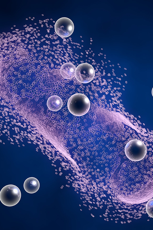

B cell malignancies originating within the skeletal compartment represent a major obstacle in clinical hematology. This specific disease arises when mature plasma cells undergo clonal expansion and malignant transformation inside the bone marrow cavity. Under normal physiological conditions, these specialized white blood cells manufacture an array of immunoglobulins necessary for adaptive immunity. However, neoplastic transformation causes these cells to secrete excessive amounts of nonfunctional monoclonal proteins, resulting in severe systemic pathology.
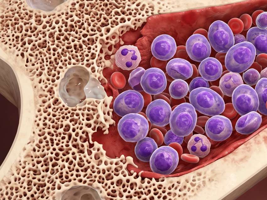
Despite the introduction of modern therapeutic combinations, including proteasome inhibitors, immunomodulatory drugs, and targeted monoclonal antibodies, long term clinical remission remains difficult to achieve. The cellular environment within the marrow protects these malignant clones, leading to inevitable disease relapse in nearly all individuals.
The pathobiological progression of this condition is governed by a distinct chain of events inside the tissue niche:
 Cytokine Mediated Survival
Stromal components secrete specialized interleukins and chemokines that constantly interact with surface receptors on the malignant cells, rendering standard therapeutic regimens ineffective.
Accelerated Osteoclast Activity
Neoplastic clones disrupt the homeostatic balance of bone remodeling, inducing severe osteolytic bone destruction, frequent fractures, and systemic hypercalcemia.
Vascular Network Expansion
Vesicles released by the tumor cells stimulate localized endothelial cells to initiate rapid angiogenesis, supplying oxygen and nutrients to the expanding clonal populations.
Immune Cell Reprogramming
Circulating regulatory factors alter local myeloid derived suppressor cells, effectively hiding the tumor from host T cell surveillance.
Consult With Our Scientists to Define Your Research Objectives
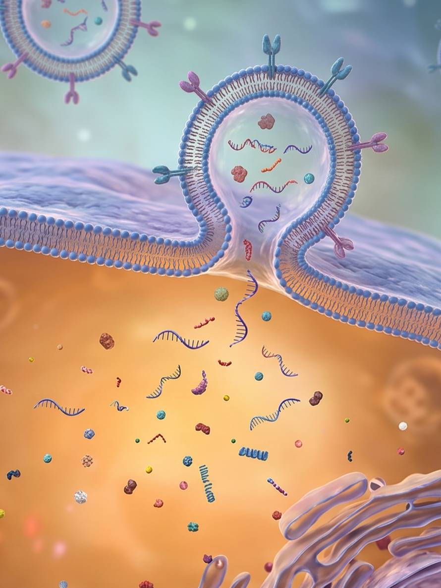
Navigating the dense and highly guarded environment of the bone marrow requires cellular carriers that can bypass host clearance mechanisms. While synthetic lipid particles face significant biological limitations, endogenous nanovesicles known as exosomes serve as highly efficient transport units. These structures are natural extracellular products involved in cellular communication, providing distinct advantages when repurposed for drug administration.
The specialized structural features of these biological vehicles include:




Engage Our Specialists to Align Your Delivery Strategy
Modern medical research focuses on utilizing these natural vehicles through two distinct functional strategies: triggering targeted cell death pathways and activating host immune responses using engineered antigens.
Tumor necrosis factor related apoptosis inducing ligand is a highly specific biological molecule capable of initiating programmed cell death in blood cancer cells without harming healthy tissue. However, direct systemic injection of raw recombinant proteins is limited by rapid kidney filtration and an exceptionally short half life. Integrating this protein into the lipid bilayer of engineered vesicles solves these stability issues.
Experimental assessments demonstrate that this structural integration yields significant benefits:
Instead of delivering direct cytotoxic agents, researchers can also configure these biological carriers to function as advanced vaccines by presenting specific disease markers to host immune cells.
| Immunotherapeutic Approach | Antigenic Components | Primary Cellular Target | Observed Physiological Outcome |
|---|---|---|---|
| Single Antigen Methods | Soluble peptide fractions | Limited cytotoxic T cell recruitment | Weak immune activation with rapid disease progression |
| Multivalent Vesicular Vaccine | Myeloma specific antigen 1 combined with Dickkopf 1 | Complete cytotoxic T lymphocyte activation | Enhanced survival rates, reduced skeletal damage, and decreased tumor volume |
Mechanistic evaluations show that co-delivering these specific markers on a single native lipid structure creates a strong, coordinated immune response, offering a viable path for personalized therapeutic design.
Discuss Your Project Specifications with Our Lead Oncology Group
Creative Biolabs maintains an advanced bioengineering platform designed to assist researchers in manufacturing and analyzing customized vesicle. Our technical services follow a sequential, step by step process to ensure maximum product purity and scientific validity.

We extract intact vesicles from various cell sources using automated tangential flow filtration,size exclusion chromatography or ultracentrifugation to remove background proteins and cellular debris.
Our facility supports diverse cargo incorporation methods, such as active electroporation for delicate nucleic acids, chemical incubation for hydrophobic molecules, and permanent donor cell modification for internal protein expression.
Every sample is documented through Nanoparticle Tracking Analysis for concentration, Transmission Electron Microscopy for structural morphology, and Western blotting to verify the presence of classic vesicle markers.
Partner With Our Research Team to Optimize Your Loading Architecture
To support accurate experimental replication and streamline development schedules, Creative Biolabs offers a standardized selection of research grade reagents manufactured under rigorous quality control standards.
Created for the fast, high yield purification of vesicles from small volume clinical serum, plasma, and cell supernatants.
Fully characterized, stable vesicle preparations obtained from verified cell lines to serve as accurate controls for sizing and quantification assays.
Membrane specific stains and conjugation reagents optimized for tracking cellular internalization kinetics and biodistribution within laboratory tissue models.
Surface-modification toolkits based on click chemistry, designed to covalently attach antibodies to vesicles while maintaining physiological reaction conditions.
Coordinate With Our Biological Experts to Design Your Study
A: Yes. We perform standard Nanoparticle Tracking Analysis on every batch post loading to provide a clear profile of particle size distribution and total concentration, ensuring the structural stability of your formulation.
A: Yes. We offer specialized fluorescent tracking tools and membrane labeling services. This allows researchers to visualize and quantify vesicle uptake kinetics within blood cancer cell lines via standard fluorescence microscopy or flow cytometry.
A: Yes. Our technical facility operates flexible culture systems that can easily scale from small-scale extraction to large-scale extraction, ensuring you receive a consistent batch of uniform quality for larger in vivo projects.
A: We perform testing for both mycoplasma and endotoxins upon request. This ensures that all engineered or isolated vesicle preparations are safe for downstream cell culture experiments and in vivo animal models.
Connect With Us to Customize Your Assay Protocol