Therapeutic Exosomes for Colorectal Cancer
Colorectal Cancer Exosome Delivery Advantages Research Our Services Available Products FAQs
Pathological Complexity and Current Barriers in Gastrointestinal Oncology
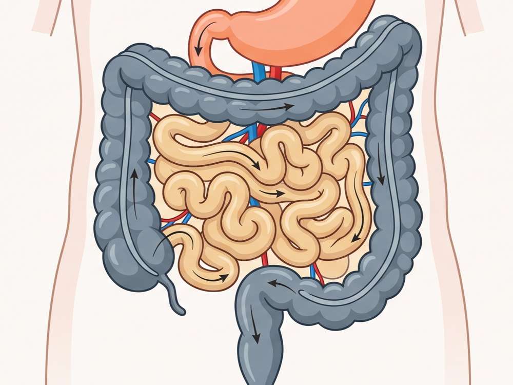
Malignant transformation within the lower gastrointestinal tract represents a significant proportion of global oncological diagnoses, with colorectal cancer (CRC) standing as a primary cause of cancer related mortality. The clinical manifestation of this malignancy involves prominent physiological disturbances, including hematochezia, persistent alterations in bowel habits, chronic abdominal pain, bloating, fatigue, secondary anemia, and rapid, unexplained weight loss. At the molecular level, the pathophysiology of this disease is characterized by systematic alterations in genomic expression, precipitating the dysregulation of multiple intracellular signaling cascades and aberrant cytokine networks that drive autonomous cellular proliferation.
While localized intervention can be effective in early stage disease, the management of advanced or recurrent colorectal malignancies faces major clinical limitations. Multi agent systemic chemotherapy regimens routinely induce drug resistance phenotypes, rendering conventional cytotoxic interventions ineffective over extended periods. Consequently, systemic metastasizing to vital organs remains the leading cause of mortality, limiting the overall five year survival rate to approximately 60%. This therapeutic barrier underscores the critical need for innovative delivery modalities capable of bypassing cellular resistance mechanisms and intercepting the intercellular communication vectors that facilitate distal tumor seeding.
Speak With Our Technical Team Regarding Your Engineering Requirements
Biophysical Suitability of Exosomes
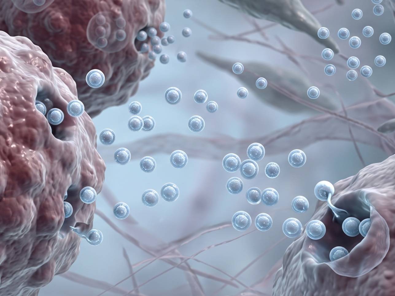
The progression and distant dissemination of colorectal malignancies depend on continuous molecular exchange between malignant epithelial cells, stromal fibroblasts, and the distant tissue niches. In this microenvironment, endogenous nanovesicles secreted by tumor cells function as critical oncogenic vehicles. These native particles transfer specific microRNAs, such as miR-21, miR-181a, and miR-23a, which directly stimulate endothelial recruitment, suppress immune surveillance, and prepare pre metastatic sites within the pulmonary, hepatic, and lymphatic systems. Serum analyses of individuals with advanced malignancies confirm a significant enrichment of vesicle bound protein markers, including Hsp60, Glypican 1, CRMP-2, Copine 3, IRF-2, and S100A9, emphasizing their systemic distribution.
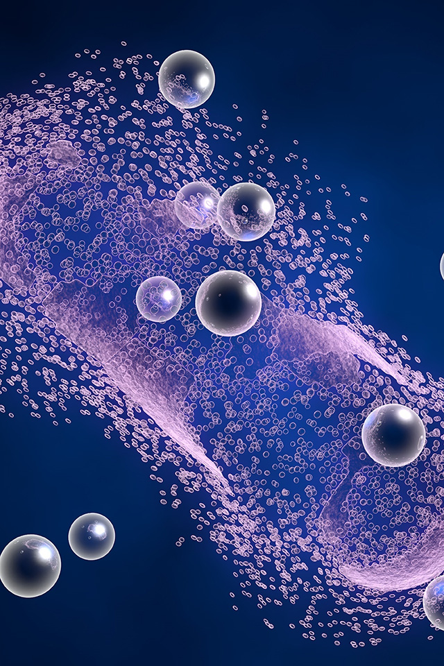
The inherent ability of these nanoscale lipid bilayers to traverse anatomical boundaries and merge with target cell membranes can be exploited to construct target specific drug delivery systems. Utilizing biological vesicles of cellular origin presents physiological advantages over conventional synthetic nanostructures:
 Direct Intestinal Epithelial Assimilation
The surface lipid composition of these natural vesicles allows for efficient integration with the intestinal mucosa, enhancing local cellular internalization without disrupting tissue integrity.
Payload Preservation
Encapsulating therapeutic candidates within the vesicular lipid bilayer shields fragile molecules from luminal enzymatic cleavage.
Low Baseline Immunogenicity
Being derived from cellular sources, these vesicle vehicles minimize systemic inflammatory signaling and avoid rapid clearance by hepatic and splenic macrophages.
Navigation Specificity
Surface proteins inherited from donor cells can guide the vesicles toward specific cellular components within the dense tumor stroma, reducing off target deposition.
Consult With Our Scientists to Define Your Colorectal Project Parameters
Exosome Delivery and Microenvironmental Reprogramming
Current research is focused on turning these natural communication networks against the tumor by utilizing donor derived or plant derived vesicles to deliver regulatory small RNAs and small molecules directly to the colorectal lesion site.
Mesenchymal Stem Cell Derived Vesicles for Signaling Interruption
Plant Derived Vesicles for Loaded Molecule Stabilization
Mesenchymal Stem Cell Derived Vesicles for Signaling Interruption
Vesicles harvested from mesenchymal stem cells (MSCs) display significant anti proliferative and protective functions in models of colorectal malignancy. These natural lipid structures can transfer specific non coding RNAs that systematically disrupt the oncogenic signaling pathways required for tumor expansion and distal migration.
Independent research groups have documented the functional mechanics of this vesicular crosstalk through distinct therapeutic angles:
-
Mesenchymal Stem Cell Vesicles: Vesicles isolated from human ulcerative colitis mesenchymal stem cells carry regulatory miR-3940-5p. This specific cargo targets and downregulates the expression of integrin alpha 6 (ITGA6), successfully restricting the growth and metastatic activity of colorectal tumor cells.
-
Bone Marrow Stem Cell Vesicles: Formulations derived from bone marrow stem cells are enriched with tumor suppressive sequences, specifically miR-22-3p and miR-16-5p. Upon internalization by malignant cells, these transcripts modulate the RAP2B and PI3K/AKT signaling cascades, arresting cell proliferation and stopping cell migration.
Plant Derived Vesicles for Loaded Molecule Stabilization
The clinical utilization of potent naturally occurring dietary compounds has been historically restricted by low aqueous solubility, poor metabolic stability, and insufficient tissue localization. For example, curcumin exhibits strong antiphlogistic and antitumor effects in laboratory models, yet delivering the active compound to colorectal sites has remained a long standing pharmacological challenge.
Recent studies show that lipid nanovesicles derived from plant sources are reliable drug delivery vehicles for the treatment of intestinal diseases. These natural structures readily incorporate large amounts of molecules, including curcumin, within their internal lipid layers. Once introduced into the gastrointestinal tract, this plant derived vesicles are internalized by intestinal epithelial cells and localized immune cells. This targeted accumulation stabilizes the compound and delivers it directly to colorectal cells. This technical approach has advanced into phase I clinical trials to evaluate safety and initial therapeutic efficacy in human subjects.
Engage Our Specialists to Align Your Custom Loading Architecture
Technical Architectures for Tailored Payload Encapsulation
Creative Biolabs has engineered an integrated technology platform to assist investigators in developing and optimizing exosome d therapies for colorectal cancer. Our solutions provide reproducible workflows across all stages of preclinical development.

We isolate clean vesicle populations from diverse matrices, such as stem cell conditioned media or plant tissues, utilizing advanced methods to remove non vesicular proteins.
Our platform supports both passive loading for compounds and active engineering methods, such as optimized electroporation or donor cell modification, for the stable packaging of regulatory oligonucleotides.
Every customized batch undergoes characterization via nanoparticle tracking analysis for size and concentration metrics, transmission electron microscopy for structural analysis, and quantitative bioassays to confirm target cell internalization.
Discuss Your Project Requirements with Our Lead Oncology Group
Analytical Control Materials and Tracking Systems
To ensure experimental reproducibility and meet the strict requirements of high impact scientific journals, Creative Biolabs offers a specialized portfolio of research grade tools manufactured under high quality standards.

Exosome Isolation Toolkits
Specialized isolation kits optimized for the clean recovery of intact vesicles from cell cultures and complex body fluid samples.

Molecular Trajectory Labels
Membrane specific fluorescent dyes and conjugation kits optimized to track the migration, uptake, and in vivo biodistribution of vesicles within gastrointestinal tissue models.

Chemical Conjugation Reagents
Advanced click chemistry reagents designed for direct covalent surface modification of vesicles, supporting antibody attachment in physiological environments.
Partner With Our Research Team to Optimize Your Loading Architecture
FAQs
Q: Can your platform support the isolation of vesicles for liquid biopsy marker validation in CRC patients?
A: Yes. We provide exosome isolation services optimized for human serum and plasma. We can isolate the vesicle fraction and perform downstream proteomic or transcriptomic screening to detect diagnostic markers such as Glypican 1, Hsp60, or specific miRNA signatures.
Q: Can you extract plant derived exosomes from sources like fruits or vegetables if I specify the material?
A: We have established extraction protocols for isolating exosomes from various plant tissues (such as ginger, grapes, or citrus fruits). You can either provide the starting plant material or have our team source it, and we will perform the sequential centrifugation and filtration steps to deliver a purified vesicle yield.
Q: Can your team test whether the vesicles are toxic to healthy intestinal cells?
A: We can run baseline cytotoxicity assays (such as CCK8 or MTT assays) on normal intestinal epithelial cell lines. This helps you determine a safe therapeutic window and ensures that the vehicle itself does not cause unintended cell death.
Q: Can you show me visual proof of the colorectal cancer cells absorbing the vesicles?
A: Yes, we can definitely do this. We can label your vesicle membranes with green or red fluorescent dyes and co culture them with colorectal cancer cell lines. Using fluorescence microscopy, we will capture clear, publication ready images showing the uptake of the vesicles inside the cells over time.
Connect With Our Staff to Customize Your Project
For Research Use Only. Cannot be used by patients.
Related Services: