Based on rich experience in antibody development, Creative Biolabs is able to offer comprehensive customized services to develop in vitro diagnostic (IVD) antibodies against different disease biomarkers, by which useful information for the cancer diagnosis, treatment, and prognosis will be obtained simply. Notably, vascular endothelial growth factor C (VEGF-C) is a potential marker for ovarian cancer and we help develop high-quality antibodies to make clients’ diagnostic tests a success.
Introduction of VEGF-C
VEGF-C, also known as Flt4-L, is a dimeric, secreted protein that belongs to the vascular endothelial growth factor/platelet-derived growth factor (VEGF/PDGF) family. In mammals, the VEGF family consists of VEGF-A, VEGF-B, VEGF-C, VEGF-D, and placental growth factor. Among them, VEGF-C shares the highest homology (30%) with VEGF-A, which is regarded as a critical angiogenic factor. This factor is expressed in many tissues, while not in peripheral blood lymphocytes.
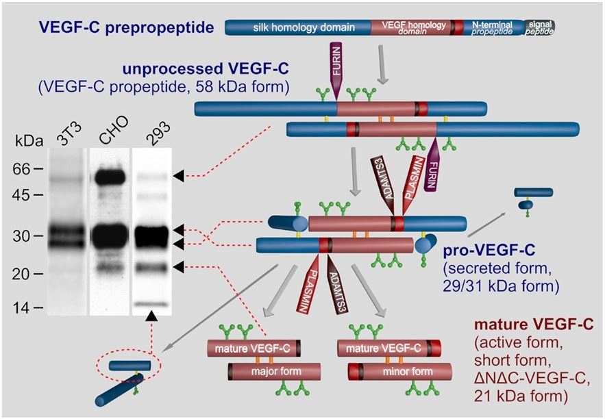
 Fig.1 Biosynthesis and activation of VEGF-C. (Rauniyar, K., 2018)
VEGF-C undergoes a complex proteolytic maturation, resulting in multiple processed forms. After translation, this molecule consists of three domains: a central VEGF homology domain (VHD), a C-terminal domain (propeptide), and an N-terminal domain (propeptide). The uncleaved VEGF-C has a size of approximately 58 kDa. The main function of VEGF-C is development- and disease-associated lymphangiogenesis, where it acts on lymphatic endothelial cells via its receptor VEGFR-3 (flt4), thereby promoting growth, survival, and migration. Apart from that, VEGF-C can facilitate the growth of blood vessels and regulate their permeability. The impact on blood vessels is mediated by its primary receptor VEGFR-3 or secondary receptor VEGFR-2 (flk1). Additionally, VEGF-C is also critical for blood pressure regulation and neural development.
VEGF-C Marker for Ovarian Cancer
In many cancers, the expression of VEGF-C is thought of important for the spread of tumor cells to lymph nodes. Tumor cells (e.g. oral squamoid, breast, and gastric cancer) expressing VEGFR-3 and VEGFR-2 can receive VEGF-C autocrine signals and activate downstream pathways that mediate aggressive phenotypes. Another source of VEGF-C, tumor-associated macrophages, contribute to the increased level of VEGF-C in microenvironments. Immune cells, such as natural killer (NK) cells, can identify VEGF-C signal and exhibit immune suppressive functions that benefit tumor progression.
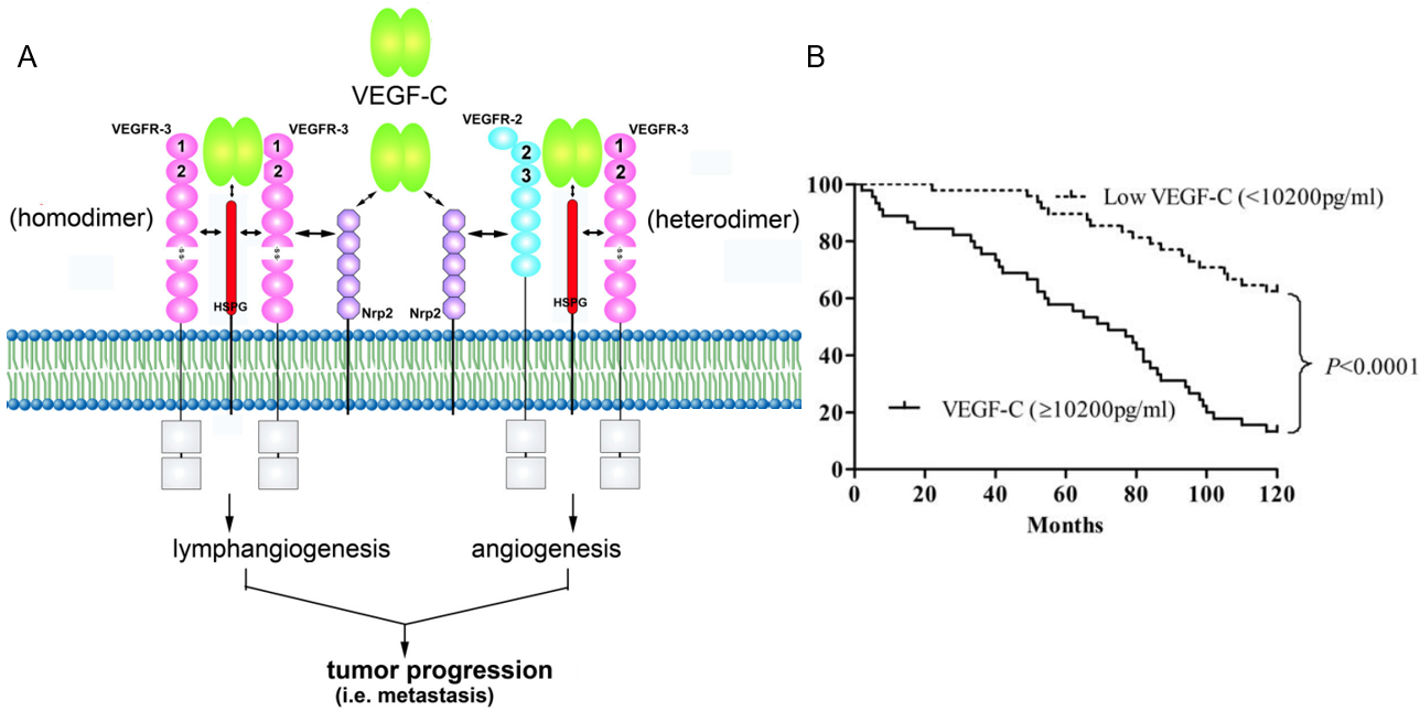
Ovarian cancer has high risk and severity in postmenopausal women and is the leading cause of gynecologic malignancy death. One report by Cheng et al. (2013) recruited 109 patients with ovarian cancer, 76 patients with benign ovarian diseases, and 50 healthy controls to determine the expression of VEGF-C. The results displayed that the serum levels of VEGF-C were significantly higher in ovarian cancer patients than those with benign ovarian diseases and healthy controls (P<0.01). Analyzed by Kaplan-Meier method, patients with high VEGF-C had remarkably shorter overall survival time than those with low VEGF-C (P<0.0001). All findings illustrated that VEGF-C may be a clinically valuable indicator for diagnostic, prognostic evaluation in ovarian cancers.
 Fig.2 (A) Functions of VEGF-C in tumor progression. (B) Kaplan-Meier survival curves. Percent survival rate was stratified by VEGF-C level.2,3
IVD Antibody Development Service for VEGF-C Marker
VEGF-C is one of the most efficient factors in regulating lymphangiogenesis. Based on the multivariate analysis and clinical parameters, serum VEGF-C has been identified as an independent adverse prognostic variable for overall survival. As a well-recognized antibody development expert, Creative Biolabs has built a novel platform for IVD antibody development against the VEGF-C marker.
Creative Biolabs has achieved a large number of challenging projects for superior antibody products and gained a great reputation from global scientists. Besides, we also provide one-stop diagnostic immunoassay development services. If you’re interested in our services, please contact us for more information or directly send us an inquiry.
References
- Rauniyar, Khushbu, Sawan Kumar Jha, and Michael Jeltsch. "Biology of vascular endothelial growth factor C in the morphogenesis of lymphatic vessels." Frontiers in bioengineering and biotechnology 6 (2018): 7. Distributed under Open Access license CC BY 4.0, without modification.
- Chen, Jui-Chieh, et al. "The role of the VEGF-C/VEGFRs axis in tumor progression and therapy." International journal of molecular sciences 14.1 (2012): 88-107. Distributed under Open Access license CC BY 3.0. The image was modified by extracting and using only part of the original image.
- Cheng, Daye, Bin Liang, and Yunhui Li. "Serum vascular endothelial growth factor (VEGF-C) as a diagnostic and prognostic marker in patients with ovarian cancer." PLoS One 8.2 (2013): e55309. Distributed under Open Access license CC BY 4.0, without modification.
For Research Use Only.