Biomarkers are essential tools for disease diagnosis, monitoring, stratification, and therapy response prediction. In vitro diagnostic (IVD) tools that are developed targeting reliable biomarkers could greatly help with these issues. As a leading service provider for antibody and immunoassay development, Creative Biolabs offers high-quality IVD antibody and immunoassay development services for different disease biomarkers, such as hector battifora mesothelial antigen-1 (HBME-1) marker.
Introduction to HBME-1
HBME-1 is a type of membrane antigen that presents in the microvilli of mesothelioma cells and other epithelial cells. It has been applied in the diagnosis of tumors stemming from the mesothelial cells. As a marker of mesothelial cells, HBME-1 has been suggested to participate in epithelial-mesenchymal transitions (EMT) linked to metastasis in neoplasms of mesenchymal origin. This antigen is useful in fine-needle aspirations and tissue specimens for thyroid malignancy, exhibiting diffuse strong stained mostly in malignant thyroid carcinomas. Thus, HBME-1 is a promising immunomarker in understanding thyroid pathology and also a universal biomarker of malignancy due to its high expression level in several aggressive tumors.
HBME-1 As A Biomarker of Follicular Adenoma
A follicular adenoma is a common, benign encapsulated tumor of the thyroid gland. It is a firm or rubbery, homogeneous, round, or oval neoplasm, surrounded by a thin fibrous capsule. The HBME-1, one of the most reliable markers in thyroid pathology, has been recognized as a critical marker of thyroid follicular origin tumors, with a higher affinity to malignant lesions compared to benign lesions.
Undoubtedly, immunohistochemistry (IHC) shows the advantage in cases where histomorphology is insufficient to establish a definitive diagnosis. The study investigated the ability of HBME-1 and other markers (CK19, S100) in differentiating between hyperplastic, benign, and malignant thyroid lesions. IHC analysis of 60 specimens (10 hyperplastic nodules, 14 follicular adenomas, and 36 malignant thyroid carcinomas) was carried out and found that HBME-1 positivity was marked in 86.1% of malignant cases while the majority of the benign lesions were negative. In addition, strong diffuse CK19 positivity confirms papillary differentiation while HBME-1 immunolabeling suggests malignancy. The utilization of markers, individual or in combination, benefits to the differential diagnosis in thyroid pathology.
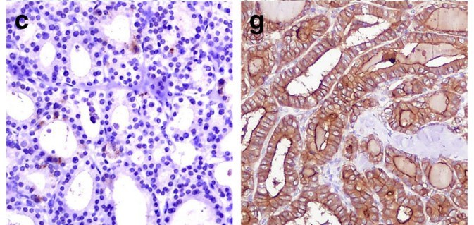 Fig.1 Expression of HBME-1 in follicular adenoma and follicular variant of papillary carcinoma (Left: absence of immunoexpression of HBME-1; Right: membranous and cytoplasmatic immunoexpression of HBME-1).1
The differential diagnosis between follicular adenoma and follicular carcinoma is difficult to determine, even with permanent sections. It is not unusual that patients diagnosed with benign follicular adenoma and then their diagnoses had to be changed to malignancies because of metastasis or recurrence. IHC staining with HBME-1 may be helpful clinically to identify cases with a high risk of recurrence in follicular carcinoma, and cases that benign adenomas require close follow-up.
IVD Antibody & Immunoassay Development Services Provided by Creative Biolabs
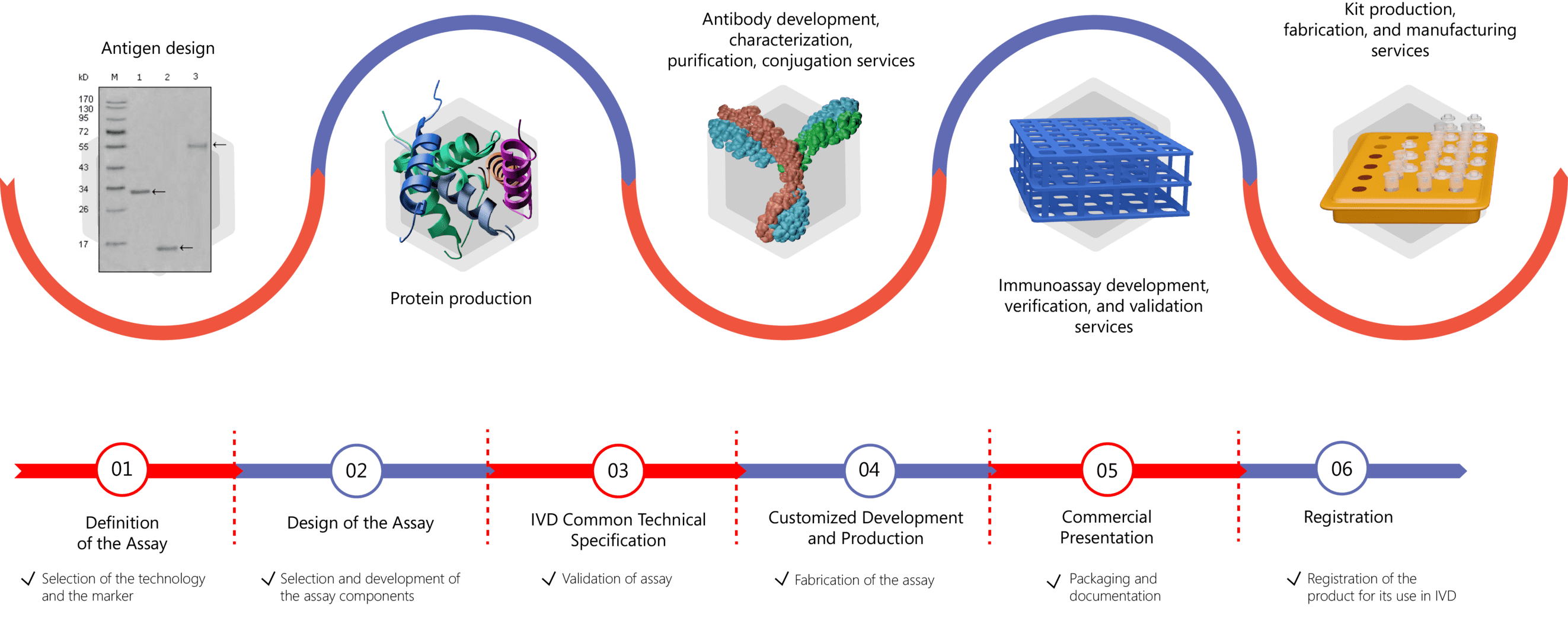
Creative Biolabs has extensive experience in contract IVD antibody & immunoassay development. We offer customized solutions for developing raw materials and immunoassays for the quantitative or qualitative detection of different disease biomarkers. We offer expertise in different phases of the kit development, including antigen design, antibody generation, recombinant protein expression, and assay development, validation, and production. For more information, please click the links below:
- IVD Antibody Development
- Antibody Pair Development
- Antibody& Protein Conjugation
- IVD Immunoassay & Kit Development
Please feel free to contact us to get more information or directly send us an inquiry.
Reference
- Dunđerović, Duško, et al. "Defining the value of CD56, CK19, Galectin 3 and HBME-1 in diagnosis of follicular cell derived lesions of thyroid with systematic review of literature." Diagnostic pathology 10.1 (2015): 1-18. Distributed under Open Access license CC BY 4.0, without modification.
For Research Use Only.