Currently, in vitro diagnostic (IVD) antibodies are increasingly used for the diagnosis and prognosis of diseases, and for drug therapy monitoring. As a leading company in the antibody development market, Creative Biolabs has established an advanced platform that allows us to offer a broad range of IVD antibody development services. Here, we introduce our high-quality IVD PPWD1-specific antibody development services.
PPWD1
Cyclophilins consist of one of the three classes of peptidylprolyl isomerases discovered in all prokaryotic and eukaryotic organisms, together with viruses. Most of the seventeen human cyclophilins with the catalytic domain are in tandem with other domains, but many particular functions of a specific cyclophilin or its relevant domains remain unclear.
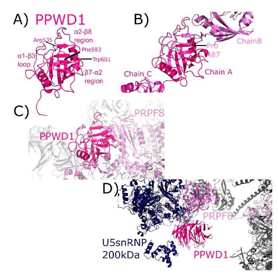
The structure of the isomerase domain from a spliceosome-correlated cyclophilin, PPWD1 which is known as peptidylprolyl isomerase domain and WD repeat containing 1 or peptidylprolyl isomerase containing WD40 repeat, has been solved to 1.65 A˚. PPWD1 acts as a protein-coding gene and its related pathways are mRNA splicing. An important paralog of this gene is PPIL2. This polypeptide encodes an N-terminal WD40 repeat domain as well as a C-terminal domain homologous to Cyps. PPWD1 was previously cloned in 1994 and subsequently purified as part of the catalytical component of the spliceosome C complex. The N-terminus of the isomerase domain in the crystal is bound to the active site of a nearby isomerase in a manner similar to the substrate. In NMR solution, this sequence binds to the active site of the cyclophilin, however, cannot be turned over by the enzyme. A pseudo-substrate N-terminus to the cyclophilin domain of PPWD1 could have wider involvements for the function on this cyclophilin in the spliceosome, where it is located in human cells.
 Fig.1 PPWD1 structures.1
Mechanisms of PPWD1 in Cancers
Gastroenteropancreatic neuroendocrine tumor (NET) commonly presents as liver metastasis from a neoplasm of unknown primary. NET is often metastasized at diagnosis. In the pilot study using cDNA microarray, scientists found gene PPWD1, CD302, and ABHD14B whose expression profiles associated with certain NET primaries. Downregulation of PPWD1 and CD302 illustrates the pancreas as a site of the primary. So scientists recently examine the gene and protein expression of the three genes in abdominal NET metastases so as to predict the location of the respective primary carcinoma.
The report elucidates that the primary NET from pancreas, stomach and small intestine along with their respective liver metastases differ from each other by the expression profile of these three genes PPWD1, CD302, and ABHB14B. The gene and protein expression of PPWD1, CD302, and ABHB14B is analyzed in abdominal NET metastases to identify the site of their respective primary carcinomas. Cryopreserved tissues from NET metastases collected from different institutions, group A (29), group B (50), and group C (132), are examined by comparative genomic hybridization. Through blindly evaluated data, gene expression analysis correctly indicates the primary in the ileum in 94% of the specimens of group A and 58% in group B. A pancreatic primary is predicted in 83% cases of group A and in 20% of group B, respectively. The combined sensitivity of group A and B is reaching 75% for ileal NET and 38% for pancreatic NET. Immunohistochemical assay with group C reveals an overall sensitivity of 80%. The gene and protein expression analysis of PPWD1 in NET metastases correctly identify the primary in the pancreas of 80% samples, provided that tissues are well preserved. Remarkably, immunohistochemical profiling demonstrates PPWD1 as the best marker for the pancreatic detection.
IVD Antibody for Pancreatic Cancer
The pancreas releases the enzymes that can help digestion, hormones and manage the blood sugar. Pancreatic cancer occurs firstly in the tissues of the pancreas, an organ in the abdomen that lies horizontally behind the lower position of the stomach. This kind of cancer typically spreads rapidly to neighboring organs, however, seldom detected in early stages. For people who have pancreatic cysts or a family history of pancreatic cancer, certain screening steps might aid to detect the problem as soon as possible. One significant sign of pancreatic cancer is diabetes, especially when it begins with weight loss, jaundice or pain in the upper abdomen that extends to the back. Treatment for the pancreatic tumor may include surgery, chemotherapy, radiation therapy or a combination of these.
Pancreatic cancer represents the vast majority, is a considerably aggressive and lethal disease with an overall five-year survival rate of only approximately 8%. It is currently the fourth leading cause of cancer mortality in both men and women. Although there are advances in understanding the tumorous biology of pancreatic cancer development, molecularly targeted therapies have not been translated into substantially improved prognosis of this deadly cancer.
Here, Creative Biolabs serves as a service provider in the in vitro diagnostic (IVD) field. With our versatile IVD platform, Creative Biolabs is proud to develop novel PPWD1-specific (paired) antibodies from scratch to commercial IVD kit (we can also start with provided antibody candidates). If you are interested in our service, please do not hesitate to contact us for more details.
Reference
- Rajiv, Caroline, and Tara L. Davis. "Structural and functional insights into human nuclear cyclophilins." Biomolecules 8.4 (2018): 161. Distributed under Open Access license CC BY 4.0, without modification.
For Research Use Only.