Coccidioides posadasii
Coccidioides Posadasii (C. posadasii) has attracted increasing attention because of its role as a causative agent of coccidioidomycosis or valley fever in humans. Absolutely, in-depth studies of C. posadasii will be propitious to the therapeutic applications of coccidioidomycosis. With abundant industry knowledge and experience, Creative Biolabs tailors quality programs and services to meet the client’s needs in the research of C. posadasii and therapeutic applications of coccidioidomycosis. Now, we focus on the introduction of C. posadasii here.
Background of Coccidioides Posadasii
C. posadasii is one member of the genus Coccidioides, and the other important pathogenic fungus is Coccidioides Immitis (C. immitis). Because of the morphological identity, it was formally known as the non-California C. immitis. However, after comparison of RFLPs within 10 DNA loci from clinical isolates of C. immitis from California, Arizona, and Texas, scientists found a significant difference among the allele frequencies, indicating C. posadasii is a separate species of Coccidioides.
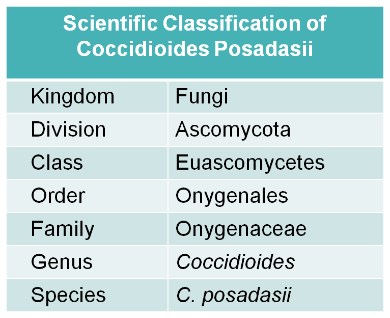 Fig.1 Scientific classification of C. posadasii.
Distribution of Coccidioides Posadasii
Morphologically identical to C. immitis, C. posadasii is present in Southwestern Unites States, Northern Mexico, as well as Central and South America. However, C. immitis is geographically largely limited to California. C. posadasii lives in the soil forming filamentous saprobes, which helps it withstand harsh conditions such as little rainfall, hot summers, low elevation, and alkaline rich soil with high salinity. It is documented that C. posadasii is capable of tolerating a pH range of pH 2 to 12.
Structure of Coccidioides Posadasii
As a dimorphic pathogen, C. posadasii has two distinct phases of development, saprobic and pathogenic. The saprobic phase is characterized by the formation of arthroconidia, which is produced by the division of hyphae and the differentiation of cellular compartments. Another typical characteristic is the up-regulated presence of the Woronin body, a protein involved in sealing septal pores in response to cell damage. The pathogenic phase is characterized by the production of mature arthroconidia and spherules, which contains endospores that can be transported by the bloodstream to other tissues of the body, particularly to the brain and central nervous system, where they can germinate and grow to cause even more severe disease. The dimorphism explains the mechanism by which C. posadasii can evade the immune system.
Coccidioides Posadasii Related Disease
As one of the most virulent of the fungal pathogens, C. posadasii causes the fungal disease coccidioidomycosis in both animal and human hosts. Beginning as a benign, inapparent or mildly severe upper respiratory infection, coccidioidomycosis occurs by the inhalation of soil containing arthroconidia formed during the mycelial phase. The parasitic phase begins when the pathogen invades the lung, then arthroconidia divide into multinucleate spherules that mature and reproduce to form endospores. Upon endosporulation, enzymatically active urease, the virulence factor, gets released and attaches to the host lung causing an inflammatory response.
Targets of Coccidioides Posadasii
β-1,3-glucanosyltransferases expressed in C. posadasii are believed to be vital for mold and yeast development, suggesting a useful target for the antibody exploration and therapeutic applications of coccidioidomycosis.
C. posadasii has become a perfect model for studying the evolutionary biology of pathogenic fungi. With several years of experience in antifungal drug discovery, Creative Biolabs spares no effort to offer valuable suggestions and reliable services for our worldwide clients to support their study. We are dedicated to partnering with you to address your research requirements. We can also offer custom services to meet your particular research needs. Please feel free to contact us for more details.
For Research Use Only.