Coccidioidomycosis
Coccidioidomycosis is a pulmonary or hematogenously spread disseminated disease caused by the fungus Coccidioides. The disease is usually acquired by inhaling spore-laden dust. As one of the top companies in antifungal drug discovery, Creative Biolabs has launched extensive and in-depth researches in the nosogenesis of coccidioidomycosis. Now, we can offer the largest and most diverse portfolio of standard and custom antifungal drug discovery services to our worldwide clients to support their antifungal studies.
Introduction of Coccidioidomycosis
Coccidioidomycosis, commonly known as cocci, Valley fever, California fever, desert rheumatism, and San Joaquin Valley fever, is a pulmonary or hematogenously spread disseminated mycotic infection. It usually occurs as an acute benign asymptomatic or self-limited respiratory infection. The most common causative agent of coccidioidomycosis is Coccidioides, which is known to live in the soil in the southwestern United States, Northern Mexico, Central, and South America. Recently, the fungus also found in parts of south-central Washington and Argentina.
There are two species of Coccidioides have been identified based on their morphological features.
-
Coccidioides Immitis
It is a pathogenic soil fungus distributed in certain parts of the southwestern United States (San Joaquin Valley of California), northern Mexico, and a few other areas in the Western Hemisphere.
-
Coccidioides Posadasii
It is endemic to certain arid-to-semiarid areas of the southwestern United States, northern portions of Mexico, and scattered areas in Central America and South America.
Types and Symptoms of Coccidioidomycosis
There are two types of coccidioidomycosis: primary coccidioidomycosis and progressive coccidioidomycosis.
-
Primary Coccidioidomycosis
People suffered from primary coccidioidomycosis are usually asymptomatic, but present nonspecific respiratory symptoms resembling those of influenza, acute bronchitis, or occasional acute pneumonia or pleural effusion. The symptoms of primary coccidioidomycosis include fever, cough, chest pain, chills, sputum production, sore throat, and hemoptysis.
-
Progressive Coccidioidomycosis
A progressive coccidioidomycosis is a long-term form of the illness, which exhibits nonspecific symptoms lasting a few weeks, months, or occasionally years. In these cases, low-grade fever, anorexia, weight loss, and weakness are typical symptoms. Sometimes, lung abscesses (infections), progressive cyanosis, dyspnea, and mucopurulent or bloody sputum may occur.
Diagnosis of Coccidioidomycosis
The diagnosis of coccidioidomycosis is different and multiple based on history and typical physical findings.
-
Serologic Testing
The blood test for anti-coccidioidal antibodies includes:
- Enzyme immunoassay, which is commonly used to diagnose coccidioidomycosis because of high sensitivity
- Immunodiffusion kit (to detect IgM or IgG antibodies)
- Complement fixation (to detect IgG antibodies)
-
Chest X-ray
It is used to check for damage to your lungs and help confirm the diagnosis.
-
Microscopic Examination
It is used to test fungal spherules in sputum, pleural fluid, CSF, exudate from draining lesions, or biopsy specimens.
-
Cultures (routine or fungal)
The culture tests on sputum (mucus you cough up from your lungs) to check for Coccidioides fungi can confirm the causative agents.
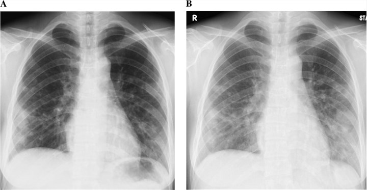 Fig. 1 Chest radiography shows enlargement of the lung nodules in coccidioidomycosis.1
Treatment of Coccidioidomycosis
The treatments of coccidioidomycosis are different based on the severity of disease.
- For mild to moderate disease, often do not require any treatment or using anti-fungal drugs.
- For severe disease, use oral and intravenous antimicrobials, especially in immunocompromised individuals.
With extensive experiences and excellent expertise, Creative Biolabs has been an optimal partner to address your research requirements in the study of coccidioidomycosis. We are very glad to advance your research and programs in antifungal agents development. Please feel free to contact us for more information.
Reference
- Lewis, E.; et al. Dust devil: the life and times of the fungus that causes valley fever. PLoS pathogens. 2015, 11(5): e1004762. Distributed under Open Access license CC BY 4.0, without modification.
For Research Use Only.