Exophiala spinifera
Exophiala spinifera (E. spinifera) is one of the etiological agents of human chromoblastomycosis, a chronic cutaneous and/or subcutaneous disease. It is important to discover the characteristics and pathogenic mechanism of E. spinifera, which is beneficial for the exploration and therapeutic applications of chromoblastomycosis. With advanced technologies and our experienced and motivated team, Creative Biolabs aims to provide our clients with efficient, reliable, and professional services in the fields of antifungal drug discovery.
Background of Exophiala Spinifera
E. spinifera is classified into the genus Exophiala, family Herpotrichiellaceae. It has been recognized as part of the black yeasts, which are a group of melanized Ascomycete fungi including species adapted to grow in humans (a wide group of human pathogens) or other hosts. As one opportunistic pathogen, E. spinifera may cause cutaneous and subcutaneous invasive infections, especially in immunocompromised and debilitated individuals. The genus Exophiala is widespread and contains about 28 species, most of which are common environmental fungi often associated with decaying wood and soil enriched with organic wastes. Some opportunistic pathogens, such as E. spinifera, E. jeanselmei, and E. dermatitidis, have been well documented.
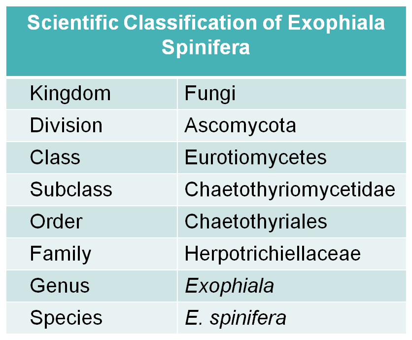 Fig.1 Scientific classification of E. spinifera.
Distribution of Exophiala Spinifera
E. spinifera has a worldwide distribution with the predomination in tropical and sub-tropical regions where the climate is warm and wet. Its common habitats include decaying wood, plants, water, and soil enriched with organic wastes. Exophiala species can also survive in the hot, humid, and oligotrophic environments such as dishwashers, steam bath facilities, and bathrooms, which are also alternative habitats for human pathogenic species. 30 °C is the optimal growth temperature of E. spinifera, but it can grow at higher temperatures such as 37 °C of the human body.
Morphological Features of Exophiala Spinifera
- The cultures of E. spinifera form mucoid, yeast-like, black colonies initially, and develop tufts of mycelium (long, brown, septate, and annellophores), finally become suede-like to downy in texture.
- Conidiophores (1-3 µm long, slightly tapering and imperceptibly annellate) are simple or branched, erect or sub-erect, spine-like with rather thick brown pigmented walls.
- Conidia are formed in basipetal succession on lateral pegs, each of which is one-celled, subhyaline, smooth, thin-walled, sub-globose to ellipsoidal. E. spinifera is characterized by toruloid hyphae and yeast-like cells with secondary.
Pathogenesis of Exophiala Spinifera
E. spinifera has been reported as an agent of cutaneous diseases, characterized by various clinical presentations of cutaneous lesions, including erythematous papules, verrucous plaques, and deep subcutaneous abscesses. The age and immune competency of the patients dictated the clinical distribution and course of diseases. It is well acknowledged that mycetoma, phaeohyphomycosis or chromoblastomycosis are the main clinical symptoms caused by E. spinifera. Occurring most commonly in tropical environments, chromoblastomycosis is a slow, progressive disease, characterized clinically by verrucous lesions, usually on the lower extremities, and appears histopathologically as sclerotic bodies in tissue. With worldwide distribution, phaeohyphomycosis usually presents in different forms in the tissues, including phaeoid yeast like cells, hyphal elements, swollen cells, pseudohyphal elements, and/or moniliform hyphae. Susceptible to multiple antimicrobial drugs in vitro, E. spinifera has been explored variable treatment modalities in the clinic.
New and effective antifungal drug discovery is crucial to preventing life-threatening fungal diseases. With extensive experience and excellent expertise, Creative Biolabs is a well-recognized research partner in the study of E. spinifera. Aided by our integrated multiple cutting-edge technologies, we can offer valuable suggestions and reliable services to meet your particular needs in therapeutic applications of fungal infection. Please feel free to contact us for more information and technical supports.
For Research Use Only.