Fonsecaea pedrosoi
Fonsecaea pedrosoi is one of the principal etiologic agents of chromoblastomycosis, a chronic fungal disease usually limited to the skin and subcutaneous tissues. Thoroughly studying the pathogenic mechanism of Fonsecaea pedrosoi is beneficial for the exploration and therapy for this fungal infection. With state-of-the-art equipment and a lot of skilled and dedicated professionals, Creative Biolabs has accumulated considerable experience in the field of antifungal drug discovery. Now we can provide best-quality products and services in a flexible and cost-effective manner in the nosogenesis and therapeutic applications of chromoblastomycosis. We focus on the introduction of Fonsecaea pedrosoi here.
Background of Fonsecaea Pedrosoi
Fonsecaea pedrosoi belongs to the family Herpotrichiellaceae. It is the predominant causative agent of Chromoblastomycosis (CBM), which is a cutaneous and subcutaneous mycosis caused by the transcutaneous inoculation of propagules (spores) from dematiaceous fungi. As a genus of ascomycetous fungi, Fonsecaea is affiliated with the family Herpotrichiellaceae and comprises three sibling species, all with pathogenic potential, namely F. pedrosoi (previously named F. compacta), F. monophora, and F. nubica.
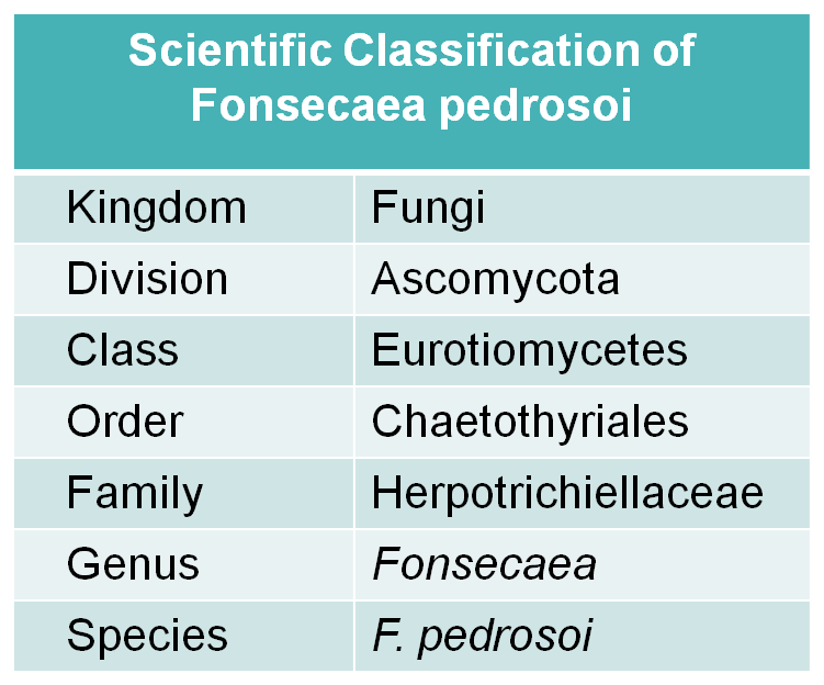 Fig.1 Scientific classification of Fonsecaea pedrosoi.
Distribution of Fonsecaea Pedrosoi
Fonsecaea pedrosoi is widely recognized as a saprophyte in the soil, rotten wood tissues, and decomposing plant materials. Although it has been found worldwide, the predominant distribution is in tropical and sub-tropical regions, especially in South- and Central America. The species F. monophora and F. nubica are closely related and they show the greater population-level genetic diversity than F. pedrosoi, which has the rigorously geographical restriction.
Physiology of Fonsecaea Pedrosoi
It is documented that the cultured Fonsecaea pedrosoi colonies are slow growing, lanose to velvety, olivaceous to black, which present growth consistently at temperatures up to 35 °C for the clinical isolates, whereas the environmental isolates exhibit growth consistently up to 35 °C, and irregularly up to 37 °C. In microscopy, Fonsecaea pedrosoi colonies are characterized by dark brown hyphae and suberect conidiophores loosely branched. Fonsecaea pedrosoi can degrade urea and tyrosine while lacking growth on the proteins gelatin, casein, xanthine, and hypoxanthine. In the life cycle, lipase activity can be tested, but phospholipase, collagenase, and amylase are not expressed.
Pathogenesis of Fonsecaea pedrosoi
Fonsecaea pedrosoi is the main agent of human chromoblastomycosis, which presents typical dermal lesions, such as Cauliflower-like nodules. And in the infected tissue, characteristic dark-colored, thick-walled, muriform cells i.e. sclerotic cells (Medlar bodies) can be observed, which has been a histopathological criterion for the diagnosis of chromoblastomycosis. Once introduced in the subcutaneous tissues, the propagules germinate to establish an invasive mycelium associated with sclerotic cells. It is notable that chromoblastomycosis is often misdiagnosed as squamous cell carcinoma in the clinic.
Antifungal Resistance
Some Fonsecaea pedrosoi strains show elevated minimal inhibitory concentrations (MICs) to amphotericin B (AMB). It is reported that Echinocandins have none or low activity against this species.
As a well-recognized research partner in antifungal drug discovery, Creative Biolabs is the best partner for you to address your research requirements in the study of Fonsecaea pedrosoi. With our integrated multiple cutting-edge technologies, we can offer valuable suggestions and reliable services for our worldwide clients to meet their particular needs. Please feel free to contact us for more details and technical support.
For Research Use Only.