
Creative Biolabs is offering the most comprehensive services for antibody development projects. With strict regulation and effective execution, we are dedicated to providing the most valuable solutions to complete your projects.
HighlightsCreative Biolabs is offering the most comprehensive services for antibody development projects. With strict regulation and effective execution, we are dedicated to providing the most valuable solutions to complete your projects.
HighlightsCreative Biolabs has established a broad range of platforms for developing novel antibodies or equivalents. These cutting-edge technologies enable our scientists to meet your demands from different aspects and tailor the most appropriate solution that contributes to the success of your projects.
HighlightsWith deep understanding in antibody-related realms and extensive project experience, Creative Biolabs offers a variety of references to help you learn more about our capacities and achievements, including infographic, flyer, case study, peer-reviewed publications, and all kinds of knowledge that can assist your projects. You are also welcome to contact us directly for more specific solutions.
HighlightsGet a real taste of Creative Biolabs, one of the most professional custom service providers in the world. We are committed to providing highly customized comprehensive solutions with the best quality to advance your projects.
Highlights Loading...
All listed services and products are For Research Use Only. Do Not use in any diagnostic or therapeutic applications.
Fibroblast growth factor 1 (FGF1, also known as acidic fibroblast growth factor) is a single-chain polypeptide with roughly 155 amino acids and molecular weight of approximately 16.5 kDa. FGF1 differs from many other secreted growth factors because it does not contain a classical N-terminal signal peptide and is released through non-conventional secretory routes under cellular stress, hypoxia or thermal injury. FGF1 interacts with high affinity transmembrane tyrosine kinase receptors (primarily FGFR1, FGFR2 and FGF4) that are complexed to heparansulfate proteoglycans which serve as necessary co-receptors upon extracellular presentation. These heterodimeric receptors cause intracellular kinase domains to autophosphorylate and then activate the RAS/MAPK, PI3K/AKT, and PLCγ signaling axes that regulate cell division, survival migratory activity differentiation. Recently discovered metabolic roles of FGF1 are powerful insulin sensitizing actions in adipose tissue and skeletal muscle that extend beyond its classical regenerative functions, demonstrated by engineered variants (e.g. S17) dissociating mitogenic function from intensive metabolic efficacy. Aberrant FGF1 signaling has been associated with abnormal angiogenesis in pathological environments such as diabetic retinopathy, tumor-tissue interactions contributing to malignant progression and chronic wounds. The multifaceted biological matter make FGF1 an attractive target for protein engineering, regenerative biopharmaceuticals and next generation metabolic therapeutics.
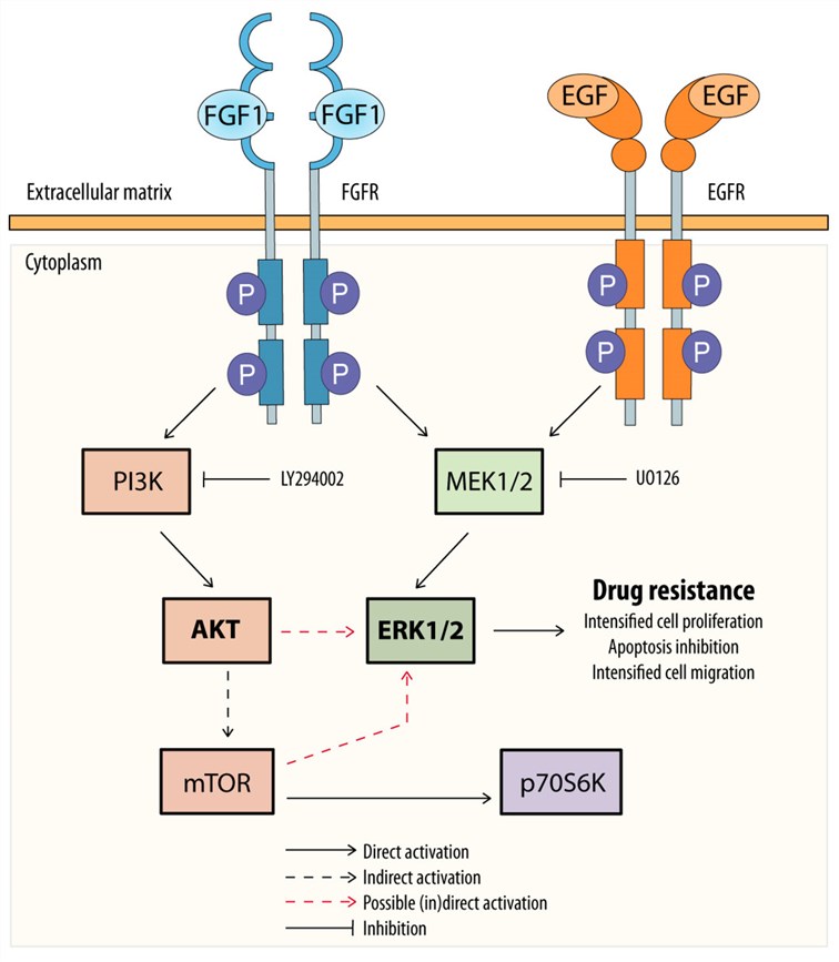 Fig.1 Potential mechanism of FGF1 and EGF action in protecting MCF-7 cells against taltobulin.1
FGF1 orchestrates a multitude of physiological programs that go well beyond its standard mitogenic activity:
We describe a focused panel of bioactive, correctly-folded FGF1 proteins engineered for the stringent demands of metabolic studies, and protein engineering. In light of the fact that genuine tertiary topology, retention of heparin-binding groove, and unaltered FGFR1/FGF2 interaction are strict requirements for both mitogenic & metabolic biological activity we employ bacterial expression systems with high intracellular solubility combined with proprietary refolding protocols catalyzing proper disulfide pairing as our production platform. YICP also provides for apparatus options including mammalian secretion systems suitable for those applications requiring useful post-translational modifications or isotope enrichment needed to carry out structural nuclear magnetic resonance (NMR). We previously developed a catalog of wild-type human FGF1, the metabolically favored S17 variant (R50V), heparin-binding enhanced mutants and cysteine-free variants to extend plasma half-life. All preparations are prepared in endotoxin-free buffers that meet the most stringent requirements for in vivo administration.
Not finding the protein product you need? Contact us to start your one-stop custom service!
To dissect its metabolic actions and facilitate discovery of therapeutics that can effectively advance regeneration, it is critical to construct cellular platforms recapitulating spatiotemporal dynamics in FGF1 secretion engaged with the cognate receptor. Through the coupling of lentiviral transduction with transposon-mediated genomic integration, we rationally developed a platform comprising multiple FGF1 stable cell lines characterized by physiologically relevant levels of expression and authentic non-conventional secretory kinetics combined with functional cellular response to stress stimuli. We previously compared constitutive versus tetracycline-inducible systems for temporal control of growth factor release, including an assortment that included metabolic biased variants like S17 mutant and co-expressions with FGF1 receptor (FGFR) pairs such as FGFR1 together with its cognate ligand. To facilitate mechanistic dissection of the cellular signaling that drives FGF1 metabolic input, we have developed reporter-coupled lines with luciferase cassettes positioned downstream of SRE, AP-1 or glucose-responsive promoter elements to enable dynamic monitoring of pathway activation during phosphokine attenuation by FGF1 stimulation or antibody blockade.
Not finding the stable cell line product you need? Contact us to start your one-stop custom service!
Advance your understanding of FGF1 with our portfolio of sequence-defined recombinant monoclonal antibodies designed to support research on this growth factor. Depending on the specific antibody and supporting datasheet, selected products may be suitable for applications such as Western Blotting, ELISA, Flow Cytometry, Immunofluorescence/Immunocytochemistry, Immunohistochemistry, and other antibody-based assays. These reagents can support the detection, localization, and analysis of FGF1 in appropriate research samples and model systems. Antibody specificity, cross-reactivity with related FGF family members, species reactivity, epitope recognition, application compatibility, and neutralizing activity, where applicable, should be determined according to the validation data available for each individual clone.
Not finding the recombinant antibody product you need? Contact us to start your one-stop custom service!
To support the above, we go beyond our generic catalog to offer fully fortified discovery and production solutions precisely aligned with FGF1-huable therapeutic & diagnostic development from natural sources:
Absolutely. Our monoclonal antibodies have been raised against unique epitopes within the FGF1 N-terminal and β-trefoil regions that exhibit minimal sequence homology with FGF2, FGF4, FGF7, FGF10, or FGF21. Extensive cross-reactivity testing by Western blot, ELISA, and cell-based neutralization confirms exquisite specificity, rendering them suitable for diagnostic quantification in complex biological matrices such as wound exudates and adipose tissue lysates.
Yes. Through optimized bicistronic and dual-promoter expression, we demonstrate coordination of FGF1 with FGFR1, FGFR2 or FGFR4 within a single stable cell line both in the presence/absence of heparan sulfate proteoglycan co-expression. Herein, we present easy-to-use stable platforms that have been functionally validated for ligand-dependent proliferation, glucose uptake and MAPK phosphorylation as ready-made cellular substrates to facilitate therapeutic screening and mechanistic dissection of receptor crosstalk.
Yes. Our bacterial expression systems and proprietary refolding protocols will yield the native β-trefoil fold, maintaining heparin-binding groove required for FGFR co-receptor assembly. The functional integrity of each batch is confirmed prior to release, through analytical verification (heparin-affinity chromatography), analysis of FGFR1 binding kinetics by surface plasmon resonance and bioactivity quantification using Ba/F3-FGFR1 cell proliferation or endothelial tube formation assays.
Certainly. These include S17 variant (R50V) to dissociate metabolic efficiency from mitogenic activity, heparin-binding enhanced mutants for extended tissue retention and site-directed substitutions designed to probe receptor subtype specificity. They are provided as purified recombinant proteins or genetically stable cell lines with complete sequence verification, mass spectrometric confirmation of identity and functional characterization by proliferation glucose uptake and heparin-affinity assays.








