
Creative Biolabs is offering the most comprehensive services for antibody development projects. With strict regulation and effective execution, we are dedicated to providing the most valuable solutions to complete your projects.
HighlightsCreative Biolabs is offering the most comprehensive services for antibody development projects. With strict regulation and effective execution, we are dedicated to providing the most valuable solutions to complete your projects.
HighlightsCreative Biolabs has established a broad range of platforms for developing novel antibodies or equivalents. These cutting-edge technologies enable our scientists to meet your demands from different aspects and tailor the most appropriate solution that contributes to the success of your projects.
HighlightsWith deep understanding in antibody-related realms and extensive project experience, Creative Biolabs offers a variety of references to help you learn more about our capacities and achievements, including infographic, flyer, case study, peer-reviewed publications, and all kinds of knowledge that can assist your projects. You are also welcome to contact us directly for more specific solutions.
HighlightsGet a real taste of Creative Biolabs, one of the most professional custom service providers in the world. We are committed to providing highly customized comprehensive solutions with the best quality to advance your projects.
Highlights Loading...
All listed services and products are For Research Use Only. Do Not use in any diagnostic or therapeutic applications.
3β hydroxysteroid dehydrogenase/steroid isomerase 1(HSD3B1, 3βHSD1) is a microsomal bifunctional integrase encoded by HSD3B1 gene, which is anchored on the membrane structure of endoplasmic reticulum and mainly expressed in skin, placenta, breast epithelium and prostate peripheral stromal cells, and has a clear tissue functional differentiation from HSD3B2, a homologous subtype specifically expressed in gonads and adrenal glands. The protein has dual catalytic activities of NAD⁺-dependent 3β hydroxysteroid dehydrogenase and steroid δ 5-δ 4 isomerase. It is a key rate-limiting enzyme that mediates the transformation of δ 5 steroid precursors into active δ 4 steroids, and can catalyze inert precursors such as pregnenolone and dehydroepiandrosterone to generate bioactive progesterone, androstenedione and testosterone. Under physiological non-gonadal conditions, basal HSD3B1 activity maintains low peripheral steroid levels to support skin homeostasis and placental hormone secretion. Under hypoxic tumor microenvironment, HIF2α-mediated transcriptional upregulation sharply elevates HSD3B1 abundance, boosting extratumoral androgen synthesis to continuously activate androgen receptor signaling and fuel castration-resistant prostate cancer growth. Its peripheral tissue-restricted steroid conversion function cannot be fully compensated by HSD3B2; germline HSD3B1 variants alter enzymatic catalytic efficiency to modify hormone tumor susceptibility, while HSD3B1 inhibition blocks intratumoral androgen production and suppresses hormone-driven carcinoma proliferation, making HSD3B1 an irreplaceable core research target for peripheral steroid enzymology and endocrine tumor therapeutic screening.
HSD3B1 is anchored in the lipid bilayer of endoplasmic reticulum membrane, which plays a dual catalytic role of dehydrogenase and isomerase. Two-step steroid modification reactions are continuously completed by relying on a single conservative active pocket, and efficient catalysis can be achieved without an independent substrate binding domain. Its unique peripheral tissue expression pattern is different from that of HSD3B2 specific to adrenal gland and gonad, which endows the body with the ability of peripheral local steroid synthesis independent of endocrine gland secretion. The steroid conversion pathway mediated by HSD3B1 can closely couple the supply of circulating inert steroid precursors with the activation of local active hormone signals, and dynamically regulate the steroid level homeostasis in peripheral tissues according to the normal physiological state and tumor microenvironment changes. This molecule is widely involved in physiological and pathological processes such as placental hormone secretion, skin lipid homeostasis maintenance and castration-resistant prostate cancer progress. The dysfunction of HSD3B1 can completely block the synthesis of Zhou Xiong hormone outside the tumor and effectively inhibit the abnormal proliferation of hormone-driven tumors. To sum up, HSD3B1 is the key target for the study of peripheral steroid enzyme mechanism and the development of targeted therapy for endocrine tumors.
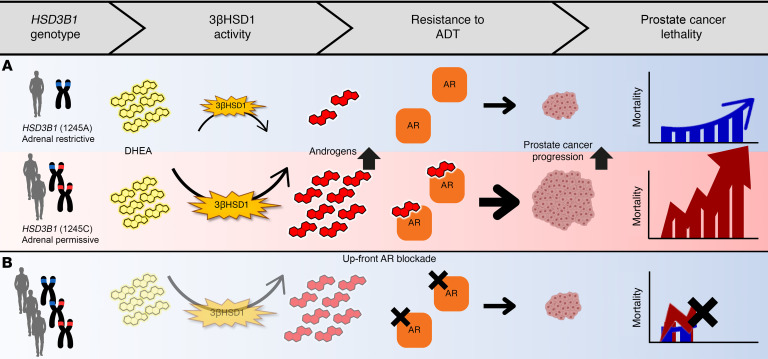 Fig. 1 Schematic diagram of HSD3B1 genotype-dependent intratumoral androgen synthesis pathway: 1245C permissive variant enhances 3βHSD1 catalytic activity to convert DHEA into androgens, driving AR activation, ADT resistance and prostate cancer lethality, while upfront AR blockade alleviates tumor progression.1
The biological functions of HSD3B1 are fully focused on dual Δ5 steroid dehydrogenation and isomerization catalytic reactions:
Creative Biolabs offers high-quality HSD3B1 proteins through optimized eukaryotic expression systems, including full-length microsomal bifunctional enzyme and isolated catalytic pocket domain variants. These products retain intact dual steroid conversion catalytic activity, suitable for steroid metabolic pathway inhibitor screening assays. All HSD3B1 proteins undergo strict quality control to ensure consistent performance and reliable application across endocrine research platforms.
Not finding the membrane protein product you need? Contact us to start your one-stop custom service!
Creative Biolabs provides custom-engineered HSD3B1 stable cell lines, including hypoxia-inducible overexpression and blank empty vector control models. These cell lines are optimized for microsomal steroid enzyme profiling and androgen receptor signal functional analysis. Each cell line undergoes stringent validation to ensure stable expression profiles during long-term hypoxic cell culture, and can be widely deployed for endocrine tumor drug screening workflows.
Not finding the stable cell line product you need? Contact us to start your one-stop custom service!
High-specificity recombinant antibodies targeting HSD3B1 are developed via advanced antibody engineering technologies, with no cross-reactivity with adrenal HSD3B2 isoform. These antibodies are validated for ER membrane localization detection and prostate/placenta tissue expression profiling, and can be combined with DHEA metabolic marker reagents to analyze peripheral steroid synthesis complexes in tumor cell models.
Not finding the recombinant antibody product you need? Contact us to start your one-stop custom service!
Beyond catalog products, Creative Biolabs offers specialized custom services for HSD3B1 research:
HSD3B1 is an ER bifunctional enzyme that converts inactive Δ5 steroid precursors into bioactive Δ4 androgens/progesterone in peripheral tissues.
Tumor HSD3B1 generates intratumoral androgens to drive castration-resistant prostate cancer, a key target for endocrine tumor therapy.
No, all HSD3B1 products and services are strictly for research use only, not intended for clinical diagnosis or treatment.
Offerings include full-length bifunctional HSD3B1 enzymes, isoform-specific detection antibodies and custom stable cell lines for steroid research.
HSD3B1 proteins are validated via dual DHEA-to-androstenedione conversion catalytic testing.








