Nowadays, in vitro diagnostic (IVD) antibody has gradually become one of the novel options presented in medical diagnosis and particularly acts as a powerful tool to dig into RCC mechanisms and then assists other therapeutics. Notably, Creative Biolabs offers high-quality antibody development services targeting numerous diagnostic biomarkers to support the IVD projects of our clients. Here, we focus on D2-40 as a marker of renal cancer.
D2-40 Marker
D2-40 is a unique monoclonal antibody directed against M2A antigen that reacts with a fixation-resistant epitope on lymphatic endothelium. M2A, also called podoplanin, consists of a 40-kDa surface sialoglycoprotein with O-linked simple mucin-type carbohydrate structure originally observed in association with germ cell neoplasms and fetal testicular gonocyte. This transmembrane protein is found to be expressed in lymphatic endothelium, epithelial cells of the choroid plexus, mesothelial cells, and osteoblasts.
D2-40 is a recently developed commercially antibody for employ on paraffin-embedded, formalin fixed tissue. In normal brains, the studies show that D2-40 is immunoreactive in ependyma, leptomeninges, subependymal, Purkinje cell layer, and white matter. D2-40 can stain tumors arising from lymphatics and certain types of vascular tumors. Interest in D2-40 initially centered on its selective immunoreactivity for lymphatic endothelium in normal tissues, since it does not stain vascular endothelium. As such, D2-40 immunostains have been proposed to improve the recognition of lymphatic invasion by primary tumors and as a marker of certain vascular lesions in clinical uses. It is valuable for this new selective marker to study benign and malignant vascular disorders in routinely processed tissue specimens. Additionally, the diagnostic utility of D2-40 in testicular and ovarian germ cell neoplasia is well-documented and it has also been proved to be a sensitive marker for seminoma metastases and primary extragonadal seminomas.
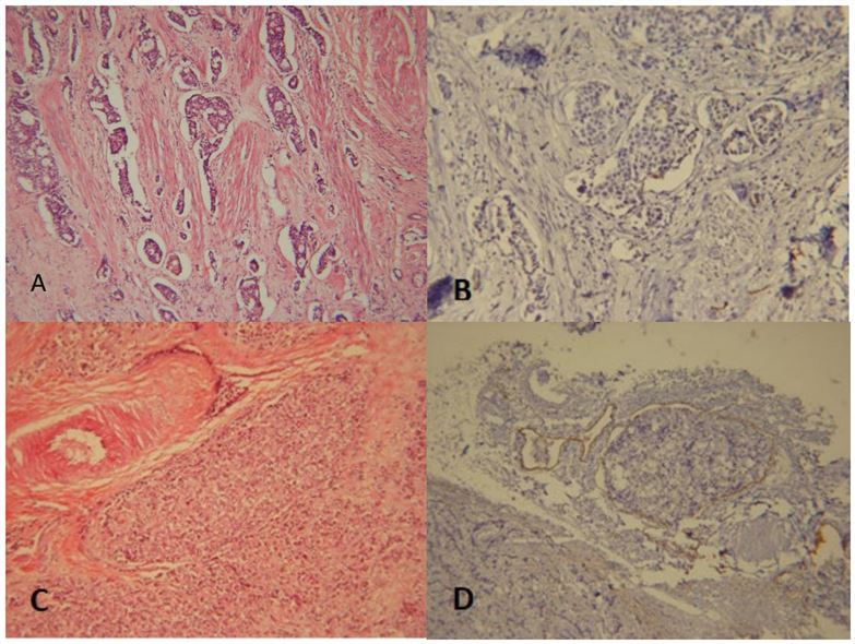 Fig.1 A. Retraction artifact (H&E method x100). B. Retraction artifact proved by D2-40 IHC (x100). C. Tumor emboli filling the lumen (H&E method x100). D. Tumor emboli proved by D2-40 IHC method (x100).1
D2-40 and Renal Cancer
It may be difficult to discriminate clear cell renal cell carcinoma (CCRCC) from neoplasms of the adrenal cortex. But this discrimination is significant in the context of possible metastasis from a CCRCC to the adrenal. Cooccurrence of solitary nodules in the adrenal gland in an RCC patient is also seen. The current established pattern of antibodies has issues and may revel variable and patchy staining. D2-40, an antibody commonly used to highlight lymphatic endothelial cells, is believed to be a definitive marker that picks op renal cortical tissue or neoplasms which can be utilized in differentiating non-neoplastic adrenal cortical cells and those of primary adrenal cortical neoplasms, from metastatic CCRCC, and from phaeochromocytoma.
IVD Antibody Development Service for D2-40 Marker
With extensive experience and advanced technologies, Creative Biolabs has been recognized as an expert in the field of IVD antibody development. Our scientific team is able to provide polyclonal, monoclonal, and recombinant antibody development services for diagnostic use. Besides, we help develop high-quality IVD immunoassays of different formats, giving expert support in feasibility analysis, protocol establishment, assay design, validation, and kit production. Our services are customized to suit the specific requirements of our clients.
If you are interested in our service, please feel free to contact us for more information and a detailed quote.
Reference
- Vosough, Zeinab, et al. "D2-40 A helpful marker in assessment of lymphatic vessel invasion in carcinoma of breast." Iranian Journal of Pathology 16.2 (2021): 96. Distributed under Open Access license CC BY 4.0, without modification.
For Research Use Only.