Tube Formation Assay
As an expert in the field of therapeutic antibody development, Creative Biolabs offers various in vitro functional assays to assess antibody bioactivity and to investigate its mechanism of action. Particularly, we offer the endothelial cell tube formation assay described here to screen antibodies with anti-angiogenic potential.
Introduction to Tube Formation & Angiogenesis
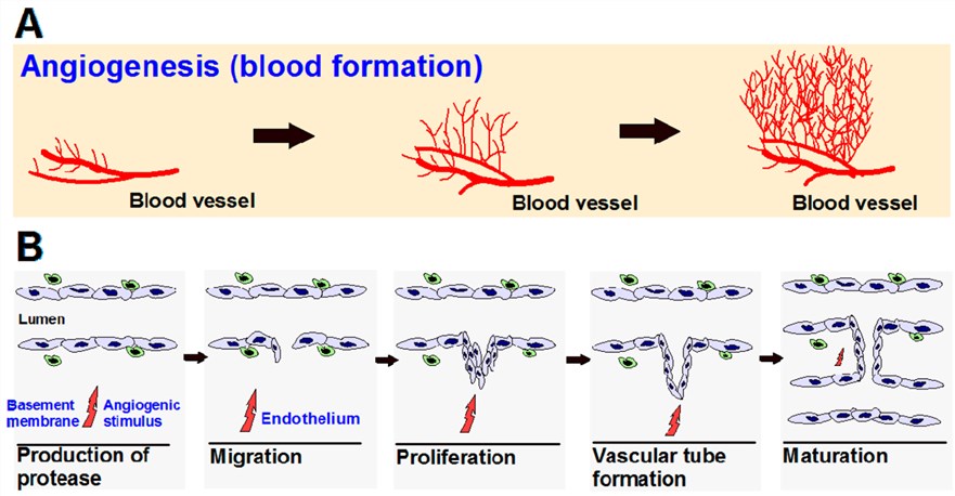
Angiogenesis, the physiological formation of new blood vessels from existing ones, is vital for various processes including organ growth, embryonic development, and wound healing. However, angiogenesis is associated with various pathological conditions, including cancer, psoriasis, diabetic retinopathy, arthritis, asthma, autoimmune disorders, infectious diseases, and atherosclerosis. Physiological angiogenesis is a highly organized process, comprising different steps: basement membrane disruption, endothelial cell migration, proliferation and differentiation into capillaries (Fig.1). One key step of this process is the assembly of endothelial cells into tubes, termed tube formation.
 Fig.1 The process of angiogenesis formulation and development.1
Tube Formation Assay at Creative Biolabs
The tube formation assay serves as a robust experimental method to assess the capacity of pharmacological agents or genetic perturbations to modulate angiogenesis, the physiological process of vascular network development. Its critical utility in identifying molecular regulators of neovascularization has established it as a cornerstone in oncology research, particularly for evaluating therapeutic targeting of pathological neovascularization.
In this assay, endothelial cells, which exhibit proliferative and migratory plasticity under angiogenic cues, are cultured on extracellular matrix substrates simulating physiological basement membranes. Exposure to pro-angiogenic factors induces cellular reorganization into three-dimensional tubular networks within hours. Quantitative parameters such as total network area, cumulative tube length, and branch node density provide standardized metrics for functional evaluation.
How to Work: End-to-end Custom Service
Step 1: Protocol Design Phase
- Jointly establish experimental objectives with collaborators, specifying investigative aims, compounds under evaluation, target cell lineages, standardized culture parameters (including replication strategy and temporal framework), and data presentation requirements.
Step 2: Preparatory Phase
- Cellular Preparation: Maintain endothelial cells in optimized culture media, expand to target quantities, and verify phenotypic integrity through viability assessments and morphological validation.
- Substrate Preparation: Reconstitute basement membrane matrix at specified concentrations, dispense into assay plates for polymerization, and validate structural homogeneity.
Step 3: Assay Implementation
- Seed cells at standardized densities onto polymerized matrices to ensure uniform adhesion.
- Administer test compounds at predefined concentrations, incorporating validated positive/negative reference agents.
- Maintain cultures under standardized incubation parameters (gas composition, thermal regulation) for protocol-defined intervals.
Step 4: Quantitative Imaging
- Acquire high-resolution micrographs of vascular networks using phase-contrast microscopy, systematically sampling multiple fields per replicate under calibrated imaging conditions. Supplemental fluorescent labeling may be employed for structural enhancement.
Step 5: Analytical Phase
- Morphometric Analysis: Quantify angiogenic parameters (network length, branch points, mesh area) via validated image analysis algorithms, ensuring analytical reproducibility.
- Statistical Evaluation: Conduct hypothesis-driven statistical comparisons with variance estimation (standard deviation, confidence intervals) to interpret compound efficacy.
- Documentation: Deliver comprehensive technical reports containing experimental schematics, methodological specifications, representative imaging datasets, quantified morphometric indices, statistical inferences, and mechanistic interpretations aligned with biological context.
Choice of Endothelial Cells
The tube-forming capacity and their response to angiogenesis stimulators vary among different groups of endothelial cells. At Creative Biolabs, we generally use the most commonly used HUVECs. Additionally, we can also use other endothelial cell types in this assay upon requests, such as HDMEC, HPMEX (Human Pleural Mesothelial Cells), and HCMEC. We can assist you in the selection of the most appropriate endothelial cell sources.
Extracellular Matrix
The extracellular matrix provides a physiological substrate that supports key cellular functions. It plays a role in the structural organization of cells and tissue, cell attachment and proliferation, as well as induction of cell differentiation. Thus, the selection of support matrices is also crucial to obtaining consistent and reliable data.
Features
- Rapid and easy to set-up
- Quantifiable and highly reproducible
- Amenable to high-throughput analysis
- Full kinetic data and automated images can be acquired
Published Data
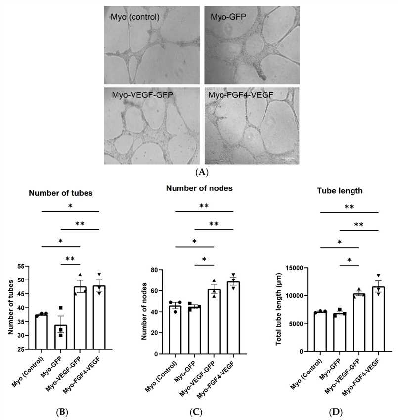
Summary: To enhance angiogenesis (new blood vessel formation), C2C12 mouse myoblast cells were engineered to overexpress human VEGF-A or both human FGF4 and VEGF-A. These cells secreted elevated levels of VEGF-A. Conditioned media from these cells promoted endothelial cell proliferation, migration, and tube formation in vitro, and increased angiogenesis in the chick chorioallantoic membrane (CAM) assay in vivo. Direct application of the engineered myoblasts on the solubilized basement membrane matrix also enhanced angiogenesis in the CAM assay.
 Fig.2 Stably transfected myoblasts' conditioned medium induces tube development in endothelial cells.2
Frequently Asked Questions
Q1: What methodology quantifies vascular network formation?
A1: Quantitative assessment employs automated image analysis algorithms to calculate morphometric parameters, including cumulative vascular network length.
Q2: How are angiogenic modulation capacities interpreted from these assays?
A2: Assay outcomes demonstrate a test agent's capacity to either enhance or suppress angiogenic processes; elevated network complexity correlates with pro-angiogenic effects, whereas diminished structural integrity indicates inhibitory activity.
The in vitro tube formation assay closely mimics the in vivo environment and is thus a powerful tool for checking angiogenesis promoters and inhibitors before using them in in vivo assays. As a leader who has been devoted to therapeutic antibody development and function analysis, Creative Biolabs takes pride in having earned a reputation for accomplishing thousands of challenging projects. Our scientists are willing to custom-design the studies to meet your specific expectations. For more detailed information, please feel free to contact us or directly sent us an inquiry.
References
- Rajabi, Mehdi, and Shaker A. Mousa. "The role of angiogenesis in cancer treatment." Biomedicines 5.2 (2017): 34. Distributed under Open Access License CC BY 4.0, without modification.
- Kennedy, Donna C., Antony M. Wheatley, and Karl JA McCullagh. "VEGF-A and FGF4 engineered C2C12 myoblasts and angiogenesis in the chick chorioallantoic membrane." Biomedicines 10.8 (2022): 1781. Distributed under Open Access License CC BY 4.0, without modification.
For Research Use Only.
