Trypanosoma brucei derived Exosome Research & Application
Overview Advantages Research Application Services Products FAQs
Biological Rationale for Trypanosome Exosome Assessment
Human trypanosomiasis, often identified as sleeping sickness, remains a severe public health challenge within Sub Saharan Africa. The etiological agent, Trypanosoma brucei, is a protozoan parasite transmitted through the bite of the tsetse fly. Upon entering the mammalian host, these parasites must navigate an intricate immune landscape to establish a persistent infection. Recent scientific evidence suggests that the survival and virulence of this pathogen are not merely functions of its motility or variant surface glycoproteins but are modulated by the secretion of extracellular vesicles. Specifically, vesicles derived from T. brucei function as sophisticated communication tools that facilitate inter parasite signaling and manipulate host physiological responses.
Consult with Our Scientists to Define Your Vesicle Validation Strategy
Functional Attributes of Trypanosoma Exosomes
Vesicles derived from the parasite exhibit unique biogenesis and compositional features that distinguish them from standard mammalian extracellular vesicles. These characteristics are central to their role in the biological success of the parasite.
Dynamic Biogenesis from Membrane Nanotubes
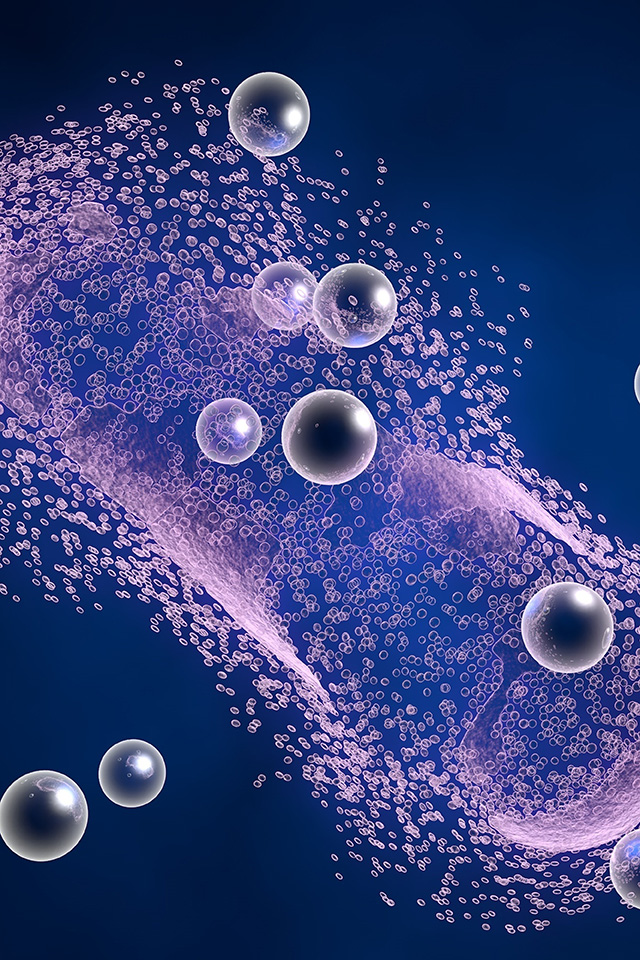
The flagellar membrane of the parasite serves as a template for the formation of elongated nanotubes. These structures subsequently fragment into spherical vesicles, increasing the surface area available for molecular exchange with the external milieu.
Enrichment of Specialized Pathogenic Proteins
Proteomic characterization reveals a high density of Variant Surface Glycoproteins and flagellar proteins within the exosomal lumen. These components contribute to the structural stability of the vesicles and function as molecular decoys during host interactions.
Specific Virulence Factor Incorporation
In subspecies such as Trypanosoma rhodesiense, the vesicles sequester serum resistance associated proteins. The horizontal transfer of these factors is essential for overcoming human innate immune defenses and establishing infectivity.
Environmentally Triggered RNA Sequestration
Under conditions of cellular stress, such as elevated temperatures or RNA processing inhibition, the vesicles incorporate Spliced Leader RNA. This cargo acts as a paracrine signal that coordinates social motility across the parasite population.
Fusion Facilitating Lipid Profiles
The exosomal membrane contains a specific concentration of phospholipids that promotes integration with host cell bilayers. This architecture is pivotal for the remodeling of host red blood cell membranes and the induction of clinical anemia.
Engage Our Researchers to Optimize Your Vesicle Analysis Protocol
Scientific Progress in Nanovesicle Mediated Communication
Research in this field has transitioned from basic morphological observation to the detailed analysis of functional outcomes in both the host and the parasite population.
Host Erythrocyte Remodeling and Anemia
A primary area of investigation involves the interaction between pathogen Nanovesicles and host red blood cells. These vesicles fuse with erythrocyte membranes, transferring parasitic lipids and proteins to the cell surface. This process alters the physical properties of the red blood cells, leading to their recognition as aberrant by the host immune system. Subsequent clearance of these remodeled cells by the spleen results in the rapid onset of anemia, a hallmark of the disease. This mechanism demonstrates that the parasite can induce distal pathology without direct presence in the tissue.
Modulation of Innate and Adaptive Immunity
Vesicles derived from the protozoan parasite exert profound effects on host mononuclear cells. Researchers have found that these vesicles establish direct communication with macrophages, inducing differentiation into both M1 and M2 phenotypes. This includes the expansion of specific subpopulations characterized by the expression of MHC I and MHC II molecules. In the adaptive immune sector, these vesicles drive the overexpression of CD3 and the nuclear factor FoxP3 in T lymphocytes. This leads to the differentiation of regulatory CD4+ and CD8+ T cells, suggesting a dual role where the parasite simultaneously promotes inflammation and suppresses targeted immune clearance to maintain a homeostatic balance during early infection.
Inter Parasite Communication and Social Motility
Beyond host interaction, these vesicles are essential for the social behavior of the parasites. In the insect host, the organism exhibits social motility, a coordinated migration pattern. The secretion of vesicles is induced by environmental stressors and the resulting vesicles are absorbed. This exchange of biological information, particularly Spliced Leader RNA, allows the population to respond collectively to stress, optimizing their migration and survival within the tsetse fly.
Discuss Your Project Requirements with Our Experts
Potentiality and Application Prospects
The unique biological roles of vesicles derived from T. brucei present several opportunities for translation.
 Diagnostic Biomarker Discovery
The presence of Variant Surface Glycoproteins and serum resistance associated proteins within circulating exosomes offers a target for noninvasive diagnostic assays. These markers could provide a more accurate reflection of the total parasite burden compared to traditional blood smears.
Acellular Vaccine Development
Given their ability to carry native parasite antigens while being non replicative, these vesicles serve as excellent candidates for vaccine research. They can stimulate both innate and adaptive arms of the immune system without the risks associated with live attenuated parasites.
Therapeutic Neutralization
Intercepting the biogenesis or uptake of these vesicles could represent a novel therapeutic strategy. For example, silencing the machinery or blocking the fusion with red blood cells could mitigate the symptoms of anemia and limit immune subversion.
Drug Delivery Systems
The natural ability of these vesicles to fuse with specific host cells can be harnessed to deliver trypanocidal agents directly to the compartments where parasites sequester, enhancing efficacy and reducing systemic toxicity.
Partner with Our Team to Refine Your Research Goals
Analytical Platforms for Exosome Development
Creative Biolabs maintains a specialized technical pipeline to support researchers in navigating the complexities of vesicles derived from parasitic sources. We provide a unified platform that covers every stage from isolation to deep functional analysis.
-
Sequential Ultracentrifugation
This remains the standard for recovering high purity vesicles from culture supernatants.
-
Tangential Flow Filtration
We utilize this technology for large scale preparation, ensuring that the biological activity of the cargo is maintained.
-
Identity Validation
Every sample undergoes characterization via Nanoparticle Tracking Analysis (NTA) for size and concentration, alongside Transmission Electron Microscopy (TEM) to confirm morphology.
-
In Vitro Cellular Assays
We assess the impact on macrophage polarization and T cell differentiation using multiparametric flow cytometry.
-
Erythrocyte Fusion Studies
Monitoring the transfer of lipids and proteins to host red blood cells to study the mechanisms of anemia.
-
Multi Omics Screening
We provide detailed analysis of the proteome, transcriptome, and lipidome within the vesicles to identify novel virulence factors or diagnostic targets.
Connect with Our Group to Advance Your Study
Specialized Reagents for Advanced Exosome Studies
To standardize exosome research, Creative Biolabs offers specialized products developed under rigorous quality control standards. These tools are designed to ensure reproducibility in scientific investigations.
Optimized for the rapid isolation of vesicles from parasite conditioned media or infected biological fluids.
Specialized dyes and kits for monitoring the internalization and trafficking of vesicles within host immune cells.
Characterized preparations to serve as positive controls in analytical and functional assays.
Engage Our Research Team to Verify Your Goal
FAQs
Q: Is it possible to evaluate the impact of these vesicles on the brain?
A: We offer specialized in vitro models to study the interaction of trypanosome vesicles with endothelial cells. This allows us to assess whether these vesicles contribute to the neurological damage and the breakdown of barrier integrity associated with the late stage of the disease.
Q: Can you provide visual proof that the vesicles are intact?
A: We perform transmission electron microscopy on isolated samples to confirm their morphology. This allows you to see the actual structure of the vesicles and ensures they remain intact and spherical after the purification process. These high resolution images are suitable for use in scientific presentations and manuscripts.
Q: Can you test how these vesicles interact with host immune cells?
A: We routinely perform coculture experiments with macrophages or T cells. Following incubation with the vesicles, we can measure changes in cell surface markers using advanced assays. This provides a direct readout of how the vesicles influence the host immune environment.
Q: What volume of culture media is required for the recovery of samples?
A: Our technical team provides guidance on the starting material needed to ensure a high yield of intact vesicles for your specific analysis.
Talk with Our Biologists to Refine Your Project
For Research Use Only. Cannot be used by patients.
Related Services: