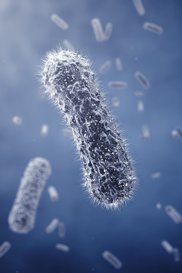

Naegleria fowleri, often termed the brain eating amoeba, is a thermophilic excavate that causes primary amoebic meningoencephalitis (PAM). This neurological infection is characterized by rapid onset and a mortality rate exceeding 97 percent. The pathogen typically thrives in warm freshwater, entering the human host through the nasal cavity before traversing the olfactory neuroepithelium to reach the central nervous system. Despite its lethality, the molecular mechanisms underlying its rapid tissue destruction and immune evasion remain inadequately understood.
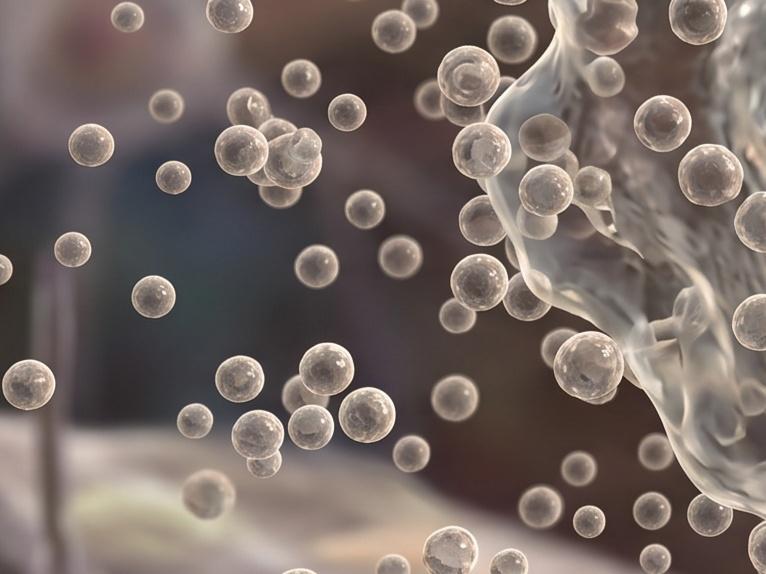
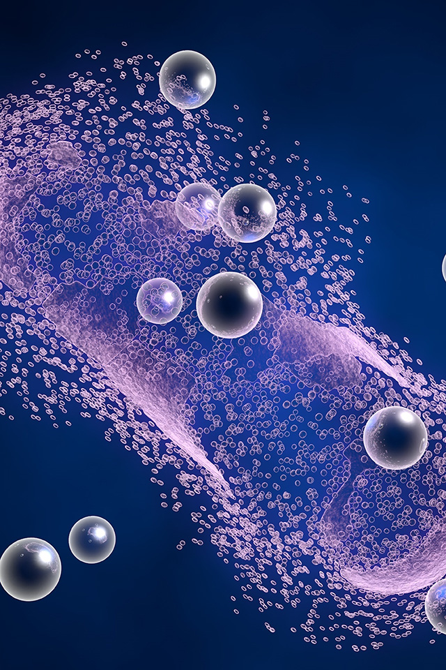
Recent scientific shifts have highlighted the role of extracellular vesicles, particularly exosomes, as critical mediators in the pathogenesis of PAM. These membrane bound nanostructures serve as distal communication tools, carrying a complex cargo of proteins, nucleic acids, and lipids from the amoeba to host cells. Investigating these vesicles is vital because they represent a contact independent mechanism of damage. By understanding how N. fowleri utilizes exosomes to prime the host environment or deactivate microglial defenses, researchers can identify novel vulnerabilities in the parasite lifecycle. This field of study is not only academic; it is a necessity for developing the rapid diagnostics and targeted interventions required to improve survival rates for this devastating disease.
Work With Our Laboratory to Design Your Custom Discovery Roadmap
Exosomes derived from N. fowleri exhibit unique biophysical and biochemical properties that distinguish them from typical mammalian vesicles. These characteristics ensure their stability and functional efficacy within the harsh and varying environments of both the external habitat and the host brain.
|
Size Heterogeneity Predominant populations range from 130 nm to 227 nm, allowing for diverse cargo capacity and tissue penetration. |
|
Proteolytic Cargo Enriched with serine proteases and cathepsin B, enabling the degradation of host extracellular matrix components. |
|
Thermal Stability Maintains structural integrity and functional activity at temperatures up to 46°C, mirroring the environmental resilience of the trophozoites. |
|
Antigenic Complexity Contains specific markers such as CD63, Hsp70, and Mp2CL5, which act as signatures for pathogen host recognition. |
|
Genetic Transport Capable of carrying specific DNA sequences (e.g., Nfa1 and NPB genes), potentially facilitating horizontal gene transfer or host modulation. |
Talk to Our Specialists to Align Your Study with Industry Standards

The scientific community has made significant strides in characterizing how N. fowleri exosomes modulate host physiology without requiring direct cellular contact. These studies utilize various cell models, including BV 2 microglial cells and THP 1 macrophages, to map out the signaling cascades triggered by vesicle internalization.
Research indicates that these vesicles are potent activators of the host immune system. Upon internalization, they trigger the upregulation of cytokines such as TNF alpha, IL 6, and IL 1 beta. This response is mediated via MyD88 dependent TLR 2 and TLR 4 pathways, leading to the activation of MAPK and NF kappa B signaling. While intended as a defense, this hyper inflammation often contributes to the catastrophic brain swelling seen in PAM.
Exposure to amoebic vesicles induces a shift in microglial cells from a homeostatic state to an amoeboid, activated morphology. This transition is accompanied by increased nitric oxide (NOS) production, further exacerbating localized tissue damage.
Beyond immune modulation, these vesicles exhibit hemolytic activity and can increase the ionic permeability of epithelial monolayers. By inducing necrosis in host cells, exosomes facilitate the paracellular invasion and migration of trophozoites through the blood brain barrier.
Mass spectrometry has revealed that the exosome cargo is surprisingly diverse, with some isolates containing over 2000 distinct proteins. Among these are leishmanolysin and other virulence factors that are instrumental in adhesion and enzymatic destruction of host tissues.
Consult Our Research Team to Address Your Complex Technical Challenges
The unique composition of N. fowleri exosomes suggests they hold immense potential for clinical translation. As the global habitat for this amoeba expands due to rising temperatures, the need for these applications becomes more urgent.
 Rapid Diagnostic Biomarkers
Given that symptoms of PAM often mimic bacterial or viral meningitis, early diagnosis is rare. Detecting specific amoebic proteins or nucleic acids within circulating or cerebrospinal fluid exosomes could provide a definitive signature for PAM in its earliest stages.
Targeted Therapeutic Interventions
Identifying the specific serine proteases or signaling molecules within the vesicles allows for the design of small molecule inhibitors. Blocking vesicle biogenesis or uptake could stall the progression of tissue damage, giving existing drugs more time to clear the pathogen.
Vaccine Scaffolds
The immunogenic nature of exosome proteins, such as the 80 KDa antigenic region recognized by polyclonal antibodies, makes them ideal candidates for vaccine research. These vesicles can act as natural delivery systems for multiple antigens, potentially conferring protective immunity in high risk populations.
Environmental Biosensors
Monitoring water sources for exosome markers or specific DNA cargo could serve as a sentinel system to predict the presence of virulent N. fowleri strains before human infection occurs.
Request a Meeting with Our Experts to Structure Your Analytical Pipeline
Creative Biolabs offers a specialized technical pipeline to support the global research community in navigating the complexities of vesicles derived from amoebae. Our platform integrates advanced biophysical techniques with deep biological insights.

We utilize high resolution methodologies to ensure the purity of isolated vesicles. Our classical protocol is sequential ultracentrifugation, followed by verification via Nanoparticle Tracking Analysis (NTA) and Transmission Electron Microscopy (TEM).
We provide granular analysis of the exosome cargo, including proteomics, transcriptomics, and lipidomics. This allows for the identification of potential virulence factors or diagnostic targets.
We assist researchers in evaluating contact independent damage mechanisms, such as membrane permeability shifts and the induction of host cell necrosis.
Collaborate With Our Staff to Advance Your Pathogenesis Investigation
In addition to our analytical services, Creative Biolabs offers specialized products designed to standardize research in the field of microorganism derived vesicles.
Inquire with Our Scientists to Enhance Your Experimental Reliability
A: Yes. Measuring the total protein content is a standard step in our workflow. We typically use BCA or similar assays to quantify the protein yield, which allows you to standardize the dosage for any subsequent functional or cell treatment experiments.
A: Yes. We use Transmission Electron Microscopy (TEM) to capture high resolution images of the isolated vesicles. This allows you to visually confirm that the exosomes maintain their characteristic bilayer structure and are free from excessive cellular debris, providing essential evidence for your research.
A: We provide comparative analysis as part of our standard service. Whether you are comparing different parasite strains or different environmental conditions (such as temperature shifts), we can provide a side by side data comparison to help you identify key differences in vesicle concentration or cargo.
A: We encourage early stage consultation. Our technical team is available to discuss your specific goals, provide advice on sample preparation, and help you select the most appropriate analytical methods. This ensures that the project design is robust and aligned with your research objectives.
Reach Out to Our Team to Finalize Your Exosome Research Framework