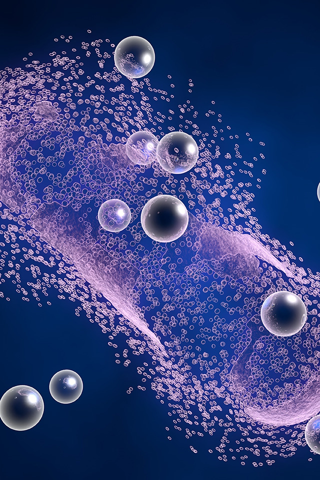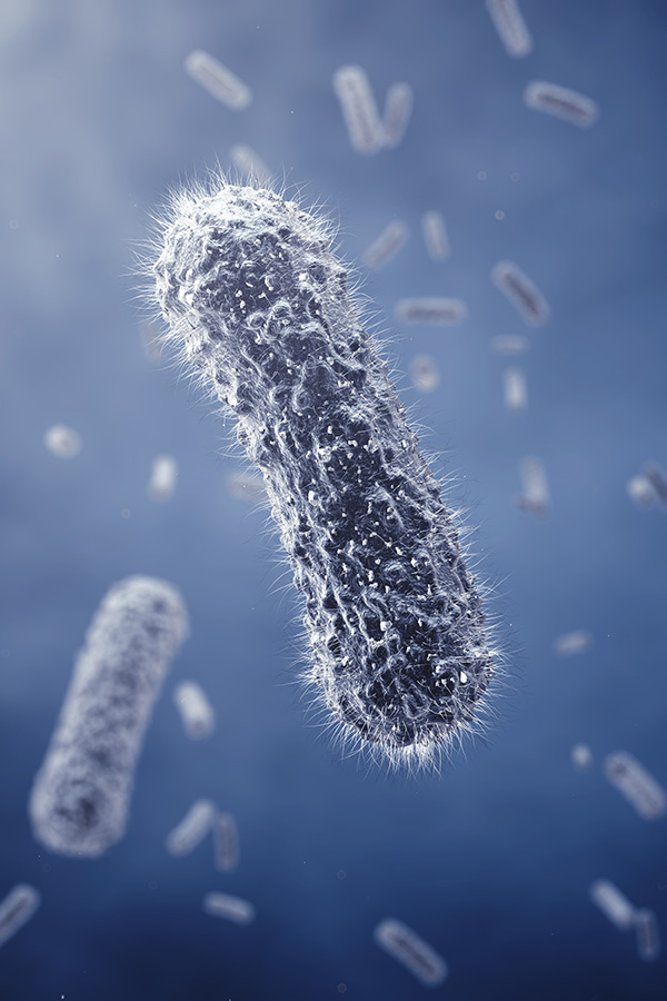

Disease caused by the protozoan parasite Trypanosoma cruzi (T. cruzi), remains a major public health challenge across Latin America and increasingly in nonendemic regions due to global migration. The biological complexity of this pathogen lies in its ability to persist within the host for decades, often leading to severe cardiac or digestive complications. Central to this persistence is a sophisticated system of intercellular communication mediated by extracellular vesicles, specifically exosomes.
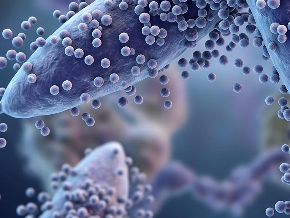
Investigation into these vesicles is essential because they function as distal effectors of the parasite. Unlike direct cell to cell contact, exosomes allow T. cruzi to manipulate host physiology from a distance, preparing a permissive environment for invasion and tempering the immune response. Deciphering the biogenesis and cargo of these vesicles provides a molecular map of how kinetoplastid parasites subvert mammalian defenses. By isolating these mechanisms, researchers can identify specific vulnerabilities in the parasite lifecycle that were previously hidden within the complexity of whole organism interactions.
Consult With Our Scientists to Define Your Research Parameters
T. cruzi exosomes possess specialized biochemical profiles and physical properties that confer distinct advantages during the infection process. These characteristics allow the parasite to maintain a high degree of environmental control.
| Feature | Functional Impact on Pathogenesis |
|---|---|
| Stability in Serum | Resistance to complement mediated lysis allows vesicles to circulate and prime distant tissues. |
| Surface Enzymatic Activity | High levels of acid and alkaline phosphatases facilitate rapid adherence to host cell membranes. |
| Selective Cargo Loading | Enrichment of MASP and GP85 family proteins ensures targeted modulation of host signaling. |
| Phase Specific Biogenesis | Differences in vesicle production between epimastigotes and trypomastigotes reflect stage specific survival needs. |
| Cytoskeletal Disruption | Ability to induce actin reorganization in host cells directly assists in parasitic internalization. |
Discuss Your Project Goals with Our Lead Exosome Researchers
Current literature has transitioned from mere identification of T. cruzi vesicles to high resolution functional analysis. Research now focuses on how these nanostructures act as vehicles for virulence factors and genomic material.
Recent studies have demonstrated that exosomes from this strain and others induce significant alterations in noninfected host cells. A primary mechanism involves the mobilization of intracellular calcium ions and changes in membrane permeability. This physiological shift not only facilitates the entry of the parasite but also leads to cell cycle arrest at the G0/G1 phase. By halting host cell division before DNA synthesis, the parasite may minimize host metabolic competition and secure resources for its own replication.
The interaction between T. cruzi vesicles and the host immune system is dualistic. On one hand, these vesicles form immune complexes with host immunoglobulin G (IgG), which can divert the immune response or trigger localized inflammation that actually assists parasitic spread. On the other hand, research involving dendritic cells (DCs) shows that vesicles can modulate the activation status of these key antigen presenting cells. Interestingly, vesicles generated during the interaction between trypomastigotes and DCs have been shown to confer a degree of resistance to lethal infection in animal models, suggesting a complex role in immune priming.
These vesicles are enriched with proteins involved in metabolism, signaling, and nucleic acid binding, confirming their role as sophisticated delivery systems for parasitic survival.
Partner With Our Scientists to Optimize Your Functional Analysis Study
The clinical and biotechnological applications of T. cruzi exosome research are vast, ranging from the development of sensitive diagnostics to novel prophylactic strategies.

 Biomarkers for Disease Monitoring
The presence of specific parasite proteins and members of the Trans-sialidase family within circulating exosomes offers a noninvasive window into the infection status. These markers are particularly valuable for monitoring therapeutic efficacy in chronic patients, where traditional blood smears often fail due to low parasitemia.
Vaccine Scaffolds
Exosomes represent a cell free therapy strategy. Because they carry native antigens in a stable, membrane bound conformation, they can stimulate potent immune responses without the risks associated with live attenuated vaccines. Studies using DC derived vesicles post interaction with the parasite indicate their potential as immunomodulatory agents that can prime the host against virulent challenges.
Targeted Drug Delivery
The natural tropism of these vesicles for specific tissues, particularly cardiac muscle and immune cells, can be harnessed. By engineering exosome mimetics or utilizing native vesicles, researchers can deliver antiparasitic compounds directly to the intracellular niche of the pathogen, reducing systemic toxicity.
Coordinate With Our Team to Tailor Your Exosome Research Strategy
Creative Biolabs provides an end to end research platform designed to address the technical hurdles inherent in studying protozoan vesicles. Our approach ensures that every stage of the exosome lifecycle is captured with high precision.

We employ advanced methodologies to separate T. cruzi vesicles from complex conditioned media or host fluids:
Our laboratory facilitates deep dives into the biological impact of isolated vesicles:
Connect With Our Experts to Accelerate Your Discovery Pipeline
To standardize research across different laboratories, Creative Biolabs offers a suite of high performance reagents specifically validated for microorganism research.
Speak With Us Regarding Your Requirements
A: We provide transcriptomic screening services to identify both small RNA and mRNA species encapsulated within the vesicles. This is crucial for understanding how the parasite might use RNA interference or other genetic mechanisms to modulate host gene expression.
A: We can utilize advanced assays to evaluate how different compounds affect exosome biogenesis or block the interaction between parasite exosomes and host cell receptors.
A: We provide Transmission Electron Microscopy (TEM) imaging services. This allows you to directly observe the characteristic cup shaped morphology and membrane integrity of the vesicles, which is essential for verifying the quality of the isolation for publication.
A: Yes. We accept samples that have already been isolated in your laboratory. Upon arrival, we perform a quality assessment to ensure the samples are suitable for specialized analysis, such as proteomic profiling or functional cell treatment assays.