
Multiple Sclerosis (MS) Modeling & Pharmacodynamics Services
Introduction
Multiple Sclerosis (MS) is a complex autoimmune and neurodegenerative disease characterized by immune cell infiltration, progressive demyelination, and axonal loss. Successful drug development requires models that can specifically isolate these components, whether it is the T-cell driven inflammation of Relapsing-Remitting MS (RRMS) or the chronic demyelination of Progressive MS. Creative Biolabs' platform provides a comprehensive suite of MS models combined with high-resolution imaging and histopathological readouts to validate your therapeutic candidates' efficacy in promoting remyelination and immunomodulation.
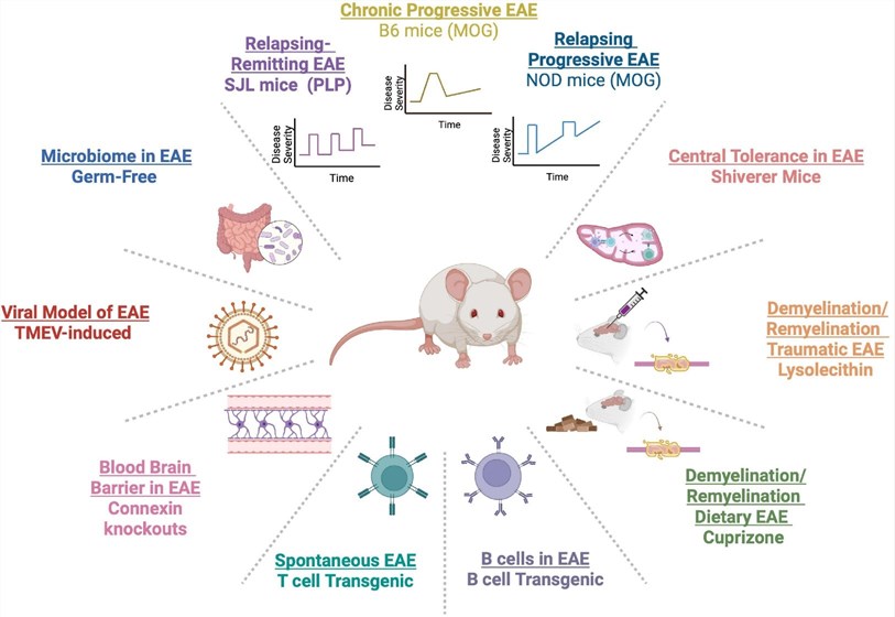 Fig.1 Various experimental autoimmune encephalomyelitis (EAE) models have been developed to simulate the diverse clinical courses and pathological features of multiple sclerosis (MS).1
Available Multiple Sclerosis Models
Creative Biolabs offers a specialized portfolio of MS models tailored to evaluate a wide range of therapeutic strategies, help you optimize drug efficacy, and delineate the Mechanism of Action (MoA) for both immunomodulatory biologics and small-molecule CNS repair therapies.
| MS Models | Modeling Method & Application Value | Animal Species |
| MOG35-55 induced EAE Mice Model | Chronic/Progressive MS. Immunization via MOG or CFA. Gold standard for long-term immunomodulators and biologics. Best for testing therapies requiring long-term immune suppression (e.g., Fingolimod, Teriflunomide, and Ocrelizumab). | Mouse |
| PLP induced EAE Mice Model | Relapsing-Remitting MS (RRMS). Immunization via Proteolipid Protein (PLP) in SJL mice. Mimics cycles of paralysis/recovery to study relapse prevention. Ideal for studying drugs that prevent relapses or maintain remission (e.g., Glatiramer acetate and Dimethyl fumarate), and rapid-acting immunomodulators and corticosteroids (e.g., Methylprednisolone), or therapies targeting Blood-Brain Barrier (BBB) integrity. | Mouse |
| MBP induced EAE Rat Model | Acute Inflammation. Lewis rats are immunized by injecting an emulsion of MBP and M. tuberculosis in IFA/PBS into the hind footpads or subcutaneously. Severe CNS inflammation with minimal or no demyelination. Highly sensitive for assessing BBB integrity and rapid anti-inflammatory responses. | Rat |
| Cuprizone induced Demyelination Model | Remyelination Focus. Selectively triggers oligodendrocyte apoptosis, leading to robust, reproducible demyelination primarily in the corpus callosum. Provides a robust, non-inflammatory platform to study oligodendrocyte apoptosis and remyelination, facilitating the evaluation of neuroregenerative agents and protein clearance therapies. | Mouse |
Evaluation Platform
Our multi-dimensional framework allows for the simultaneous assessment of immune modulation and structural repair:
-
Functional & Behavioral Readouts:
- EAE Clinical Scoring: Daily standardized scoring of tail and limb paralysis (0–5 scale).
- Motor Coordination: Rotarod and automated gait analysis to detect subtle locomotor deficits during remission or progression.
-
In Vivo Neuroimaging:
- MRI (T2 & DTI): Longitudinal tracking of lesion volume and white matter integrity (Fractional Anisotropy).
- Optical Coherence Tomography (OCT): Measuring Retinal Nerve Fiber Layer (RNFL) thickness to assess optic neuritis and neurodegeneration.
-
Histopathology & Ultrastructure:
- Luxol Fast Blue (LFB): Classic staining to quantify total demyelinated areas.
- Immunohistochemistry (IHC): Quantifying MBP (myelin), Iba1 (microglia), CD4/CD8 (T-cells), and Olig2 (oligodendrocytes).
- Electron Microscopy (EM): The "Gold Standard" for measuring G-ratio and myelin sheath thickness.
Applications
- Immunomodulator Screening: Testing the ability to reduce experimental autoimmune encephalomyelitis (EAE) clinical scores and CNS leukocyte infiltration.
- Promyelinating Therapy Validation: Assessing the differentiation of oligodendrocyte precursor cells (OPCs) into mature myelinating cells in Cuprizone models.
- Neuroprotection Studies: Investigating the preservation of axonal integrity independent of inflammatory suppression.
- Blood-Brain Barrier (BBB) Integrity: Evaluating drug impact on BBB leakage using Evans Blue or MRI contrast agents.
Our Advantages
- Phase-Specific Modeling: Ability to target specific MS phenotypes (inflammatory vs. demyelinating).
- High-Resolution Histology: State-of-the-art IHC and EM services to provide definitive evidence of remyelination.
- Longitudinal Data: Non-invasive monitoring (MRI/OCT) reduces animal numbers and provides human-relevant data.
- Expert Interpretation: Data analyzed by neuropathologists specializing in glial cell biology.
Work with Us
- Summarize the project requirements and fill in the information collection form.
- Sign a CDA from both parties to further communicate information, such as targets.
- Select an animal model, discuss experimental design, and determine assay parameters.
- Project costing and project schedule forecasting.
- We provide a detailed project plan, including the required sample quantities, methods, and protocols.
- Both parties confirm the project details and start the project.
- Confirm the timeline of the project.
- We provide periodic results and information on the animal's condition.
- We will work together to make project adjustments as necessary.
- We provide a comprehensive project report promptly.
- We arrange transportation for the produced samples.
- We provide a discussion of the project results and help to arrange the next steps.
- Data storage and archiving.
FAQs
-
Q: Can EAE results predict human clinical success?
A: EAE is excellent for predicting anti-inflammatory efficacy (e.g., B-cell/T-cell blockers) but less effective for primary progressive MS, where we recommend the TMEV or chronic Cuprizone models.
-
Q: Which MS model is most suitable for testing a drug that targets the Relapsing-Remitting phase?
A: The PLP induced EAE model (SJL mice) is the most appropriate. Unlike the chronic MOG model, PLP induced EAE exhibits distinct waves of paralysis followed by partial recovery (remission), mimicking the clinical course of RRMS. This allows for the evaluation of drugs intended to prevent relapses or extend remission periods.
-
Q: How do you differentiate between an "immunomodulatory" effect and a "pro-myelinating" effect?
A: We utilize the Cuprizone model to isolate pro-myelinating effects. Because Cuprizone induced demyelination is a toxin-driven process that occurs independently of T-cell infiltration, any improvement in myelin density can be attributed to direct effects on oligodendrocytes or OPC differentiation, rather than general immunosuppression.
-
Q: Can you assess BBB integrity in your EAE models?
A: Yes. We use Evans Blue extravasation or Sodium Fluorescein (NaF) assays to quantify BBB leakage. Additionally, we can perform high-field MRI (T1-weighted with Gadolinium contrast) to visualize and track active lesion areas where the BBB has been compromised in vivo.
-
Q: What is the standard duration for a MOG induced EAE study?
A: A typical study lasts 28 to 40 days. Immunization occurs on Day 0, with clinical symptoms (tail atony) usually appearing between Days 9 and 14. We generally track the animals until the disease reaches a stable chronic plateau to assess the long-term neuroprotective effects of the candidate drug.
-
Q: Do you offer Neurofilament Light Chain (NfL) as a biomarker?
A: Absolutely. NfL is our primary translational biomarker for axonal damage. We use high-sensitivity assays (such as SIMOA or MSD) to detect NfL levels in plasma or CSF, providing a quantitative bridge between our preclinical results and clinical trial readouts.
-
Q: How is the clinical scoring performed to ensure data integrity?
A: All clinical scoring (0–5 scale) is performed by blinded observers who are unaware of the treatment groups. Animals are scored daily at the same time to account for circadian variations, ensuring high reproducibility and minimizing bias.
-
Q: Can you perform EM for myelin analysis?
A: Yes. While LFB is excellent for screening, Transmission Electron Microscopy (TEM) is the gold standard we use to calculate the G-ratio (the ratio of the inner axonal diameter to the total outer diameter). This provides definitive proof of the thickness and quality of the new myelin sheaths formed during remyelination.
Published Data
Treatment with 670 nm light significantly upregulates the expression of the anti-apoptotic gene Bcl-2 and increases the Bcl-2: Bax ratio in the spinal cord of female C57BL/6 mice with MOG35-55 induced EAE. Furthermore, TUNEL staining confirms a significant reduction in the number of apoptotic cells across all disease stages, demonstrating the potent anti-apoptotic effect of 670 nm photobiomodulation within the central nervous system.
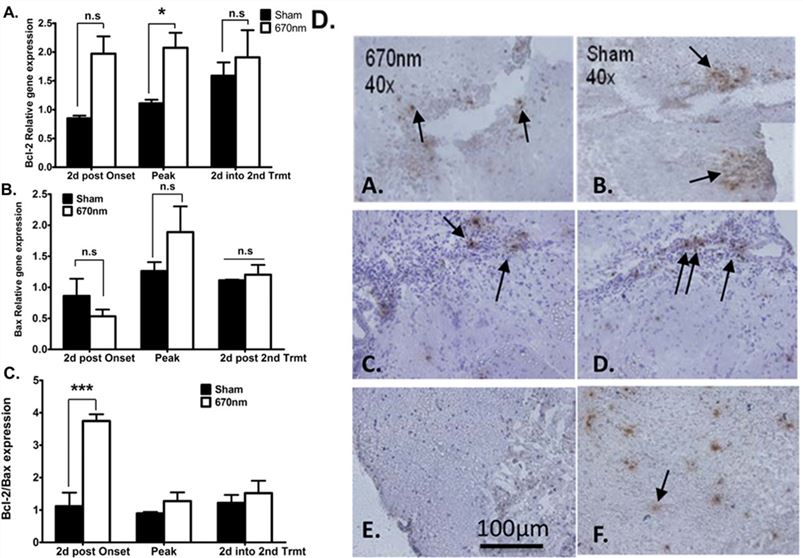 Fig. 2 670 nm light regulates apoptosis in EAE mice.2
References
- Melamed, Esther et al. "Advantages and limitations of experimental autoimmune encephalomyelitis in breaking down the role of the gut microbiome in multiple sclerosis." Frontiers in Molecular Neuroscience vol. 15 1019877. Distributed under Open Access license CC BY 4.0, without modification. https://doi.org/10.3389/fnmol.2022.1019877
- Muili, Kamaldeen A et al. "Photobiomodulation induced by 670 nm light ameliorates MOG35-55 induced EAE in female C57BL/6 mice: a role for remediation of nitrosative stress." PLOS ONE vol. 8,6 e67358. Distributed under Open Access license CC BY 4.0, with modification. https://doi.org/10.1371/journal.pone.0067358
For Research Use Only.