
LPS induced Neuroinflammation Modeling & Pharmacodynamics Service
Introduction
Neuroinflammation is a pivotal driver in the onset and progression of various neurodegenerative and psychiatric disorders, including Alzheimer's disease (AD), Parkinson's disease, Huntington's disease (HD), Multiple sclerosis (MS), Amyotrophic lateral sclerosis (ALS), and Major Depressive Disorder (MDD). Central to this process is the innate immune system of the Central Nervous System (CNS), where microglia act as the primary responders. While microglia normally maintain CNS homeostasis by scavenging plaques and pruning synapses, their chronic or irregular activation triggers a self-perpetuating cycle of neurotoxicity. This pathological state leads to the sustained release of pro-inflammatory mediators, ultimately resulting in neuronal apoptosis, synaptic degeneration, and profound cognitive dysfunction. To meet the rigorous demands of modern drug discovery, Creative Biolabs utilizes Lipopolysaccharide (LPS), a potent endotoxin and TLR4 agonist, to induce a robust, highly reproducible neuroinflammatory response that provides a high-fidelity simulation of rapid microglial activation and the subsequent "cytokine storm." This platform establishes a critical experimental window for evaluating the pharmacodynamics and efficacy of novel anti-inflammatory candidates (e.g., NLRP3 inhibitors and TLR4 antagonists), neuroprotective agents focused on preserving synaptic integrity and neuronal survival, and glial modulators designed to shift microglia from a pro-inflammatory to a neuroprotective phenotype. By bridging the gap between acute immune activation and chronic neurodegeneration, our platform delivers the scientific rigor and high-resolution data necessary to accelerate your neuroprotective pipeline.
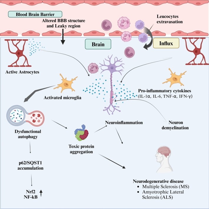 Fig.1 Compromised tight junction structures in the blood–brain barrier (BBB) cause peripheral immune cell infiltration and leucocyte extravasation, resulting in inflammation.1
Available LPS induced Neuroinflammation Models
Our platform offers a versatile range of delivery routes to precisely tailor inflammatory profiles to your specific research objectives: systemic challenges (IP/IV) are utilized to model systemic-to-central inflammation and BBB disruption accompanied by "sickness behavior," while intracerebroventricular (ICV) injections bypass the blood-brain barrier to trigger acute, localized neuroinflammation without systemic interference. For disease-specific research, we provide precision intrastriatal or intranigral microinjections to replicate localized neuronal loss relevant to Parkinson's and Alzheimer's pathology, all supported by standardized, high-reproducibility protocols in both mice and rats.
| LPS induced Neuroinflammation Models | Modeling & Application Values | Animal Species |
| Systemic LPS Model | IP or IV administration of LPS. The Systemic LPS Model investigates peripheral-to-CNS signaling, modeling sepsis-associated encephalopathy and BBB disruption caused by systemic cytokine surges. It serves as a high-translational platform for evaluating anti-cytokine storm agents and BBB-protective compounds by correlating systemic insults with "sickness behavior" and neuroinflammatory progression. | Mouse, Rat |
| Central LPS Model (ICV) | ICV injection via stereotaxic surgery. The ICV bypasses the BBB to isolate primary CNS inflammation, triggering direct glial activation and synapse loss without systemic interference. It is ideal for simulating NMDAR-related cognitive deficits and evaluating TLR4 antagonists, CNS-penetrant small molecules, and glial modulators aimed at restoring neurohomeostasis. | Mouse, Rat |
| Site-Specific LPS Model | Stereotaxic microinjection into targeted regions (e.g., Hippocampus, Striatum). The Site-Specific LPS Model focuses on localized neurodegeneration by replicating neuronal loss and circuitry disruption in regions like the substantia nigra or hippocampus. Highly relevant for Parkinson's disease and AD research, it provides a precision platform for evaluating NLRP3 inhibitors, neuroprotective agents, and anti-pyroptosis candidates designed to halt regional damage and preserve motor or cognitive functions. | Mouse, Rat |
Evaluation Platform
Our platform provides an end-to-end analytical framework to evaluate the pharmacodynamics of anti-inflammatory and neuroprotective candidates. By combining molecular profiling with functional readouts, we deliver a holistic view of how your compound modulates the LPS-induced neuroinflammatory cascade.
- Neuroinflammatory Biomarker Profiling: We provide high-sensitivity quantification of key inflammatory mediators across the brain and periphery, featuring multiplexed analysis of pro-inflammatory cytokines (e.g., IL-1β, IL-6, TNF-α, and IFN-γ) via ELISA or MSD, chemokine tracking of MCP-1 and MIP-1α to assess immune cell infiltration, and broad-spectrum oxidative stress profiling (e.g., ROS/RNS, SOD, and MDA levels) to evaluate secondary tissue damage.
- Advanced Neuroimaging & Histopathology: We visualize the cellular response to inflammation with high-resolution morphology by utilizing quantitative IHC/IF for Iba1 and GFAP, including skeletal analysis to characterize the transition of microglia and astrocytes from resting to activated states, alongside BBB integrity assessments via tight junction protein expression (e.g., Claudin-5, Occludin) and Evans Blue (EB) extravasation assays, and neuronal integrity evaluations using Nissl staining and NeuN quantification to measure inflammation-induced neuronal loss.
- Behavioral & Functional Phenotyping: We translate molecular changes into clinically relevant functional insights by integrating sickness behavior tracking (monitoring lethargy, anhedonia via sucrose preference, and locomotor activity) with cognitive impairment assays, such as the Y-Maze and Morris Water Maze, to evaluate neuroinflammation-driven memory deficits, while utilizing in vivo neurophysiology (EEG/LFP monitoring) to detect critical disruptions in network oscillations and synaptic transmission.
- Target-Specific Mechanism of Action (MoA): We provide an in-depth investigation into MoA by analyzing the TLR4 signaling pathway, specifically through the quantification of NF-κB translocation and MAPK (e.g., p38, JNK) phosphorylation, alongside an in-depth evaluation of inflammasome activation focusing on NLRP3 assembly and Caspase-1 cleavage.
Applications
- Anti-inflammatory Screening: Rapid evaluation of candidates for their ability to suppress pro-inflammatory cytokine release.
- Neuroprotective Validation: Testing drugs aimed at preventing inflammation-induced neuronal apoptosis or synaptic loss.
- BBB Research: Assessing the efficacy of compounds in maintaining or restoring BBB structural integrity.
- Mechanism of Action (MoA): Investigating the modulation of TLR4, NF-κB, or NLRP3 inflammasome pathways.
Our Advantages
- Targeted Delivery Routes: We offer systemic (IP/IV) or localized (ICV) LPS administration to mimic either systemic-derived neuroinflammation or direct CNS insults.
- Acute vs. Chronic Modeling: Tailored dosing regimens to simulate the transition from acute cytokine storms to chronic neurodegenerative-like states.
- Strain-Specific Customization: Validated protocols across multiple B6 and SD backgrounds to account for genetic variability in immune responses.
- Deep Phenotyping Accuracy: Utilization of AI-driven behavioral tracking to differentiate between general sickness and specific cognitive or social deficits.
- High-Fidelity Replication: Standardized LPS protocols that achieve highly reproducible inflammatory baselines across study cohorts.
- Translational Biomarkers: Integration of ultra-sensitive biochemistry to correlate behavioral recovery with brain-specific cytokine levels.
- Controlled Environment: Studies conducted in sound-attenuated suites to minimize stress-induced confounding of inflammatory readouts.
Work with Us
- Summarize the project requirements and fill in the information collection form.
- Sign a CDA from both parties to further communicate information, such as targets.
- Select an animal model, discuss experimental design, and determine assay parameters.
- Project costing and project schedule forecasting.
- We provide a detailed project plan, including the required sample quantities, methods, and protocols.
- Both parties confirm the project details and start the project.
- Confirm the timeline of the project.
- We provide periodic results and information on the animal's condition.
- We will work together to make project adjustments as necessary.
- We provide a comprehensive project report promptly.
- We arrange transportation for the produced samples.
- We provide a discussion of the project results and help to arrange the next steps.
- Data storage and archiving.
FAQs
-
Q: What is the peak window for measuring neuroinflammation after LPS injection?
A: Pro-inflammatory cytokines typically peak between 2–6 hours post-injection, while microglial morphological changes are most prominent at 24 hours.
-
Q: Can this model be used for chronic neuroinflammation studies?
A: While LPS is primarily an acute model, we can perform repeated low-dose injections to simulate a sustained inflammatory state.
-
Q: Do you offer positive controls?
A: Yes, we routinely utilize Dexamethasone or Minocycline as reference compounds to validate anti-inflammatory responses.
Published Data
In mouse models, intraperitoneal pretreatment with Amlexanox specifically inhibits LPS-induced IKKε expression in brain tissue, leading to a significant reduction in pro-inflammatory cytokines such as TNF-α, IL-1β, and IL-6 in both the brain and serum. By suppressing microglial activation, Amlexanox alleviates neuroinflammation, providing robust in vivo evidence for its potential as a therapeutic agent for neuroinflammatory disorders.
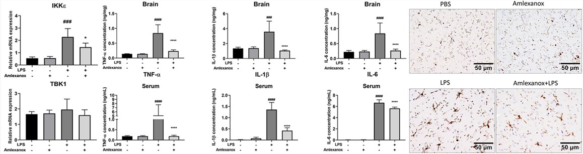 Fig. 2 Amlexanox alleviated LPS-induced neuroinflammation in vivo.2
References
- Tamatta, Rajesh et al. "Neuroinflammaging and the Immune Landscape: The Role of Autophagy and Senescence in Aging Brain." Biogerontology vol. 26,2 52. Distributed under Open Access license CC BY 4.0, without modification. https://doi.org/10.1007/s10522-025-10199-x
- Phan Van, Thach et al. "Amlexanox attenuates LPS-induced neuroinflammatory responses in microglial cells via inhibition of NF-κB and STAT3 signaling pathways." Scientific Reports vol. 14,1 2744. Distributed under Open Access license CC BY 4.0, with modification. https://doi.org/10.1038/s41598-024-53235-5
For Research Use Only.