
Spinal Cord Injury Modeling & Pharmacodynamics Service
Introduction
Spinal Cord Injury (SCI) is a catastrophic neurological event with a profound global impact; according to the World Health Organization, between 250,000 and 500,000 people suffer a spinal cord injury every year, often resulting in permanent disability and significant socioeconomic burdens. The condition triggers a complex cascade of primary mechanical damage followed by secondary neuroinflammation, axonal retraction, and the formation of a dense glial scar. Developing effective therapies, ranging from neuroprotective small molecules to regenerative stem cell and gene therapies, requires high-fidelity models that replicate these intricate pathological phases. Creative Biolabs' platform offers a comprehensive suite of SCI models combined with advanced locomotor kinematics and histological quantification to provide the rigorous evidence needed to accelerate your candidate's path to clinical validation.
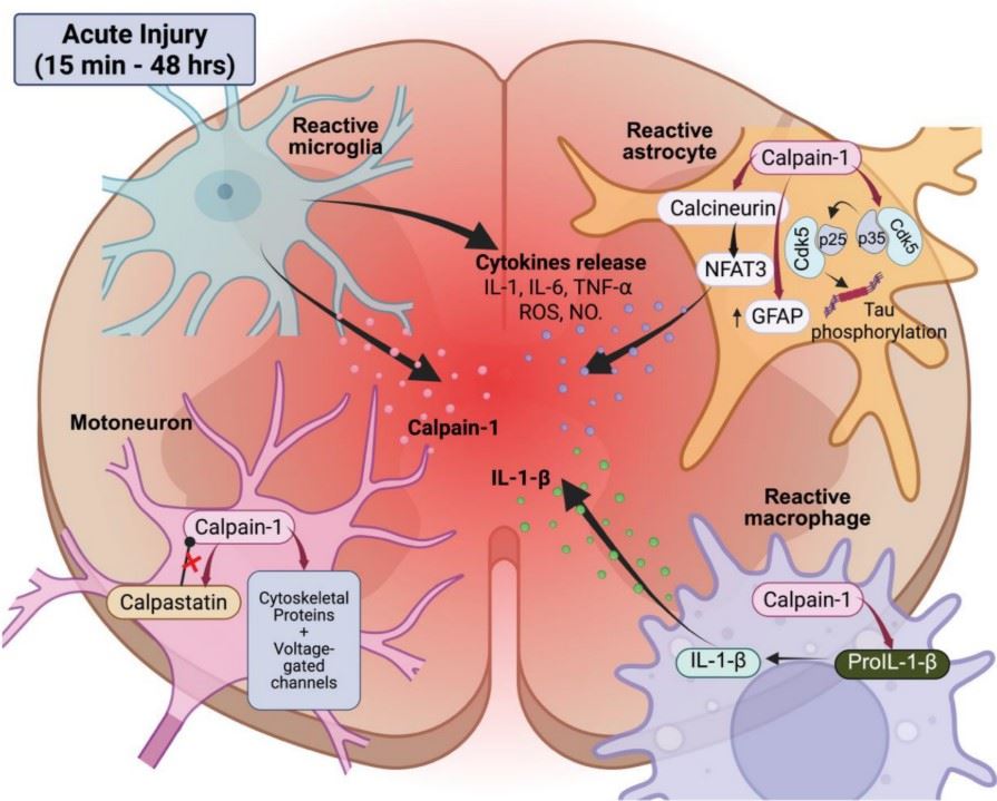 Fig.1 Acute calpain activation and neuroinflammatory responses in the spinal cord after injury.1
Available Spinal Cord Injury Models
We provide a comprehensive Spinal Cord Injury (SCI) research platform featuring precision contusion, compression, and transection models to accelerate preclinical drug development. Our capabilities span both Rodent and non-Human Primates (NHPs) models, even the Primate Peripheral Nerve Injury (PNI) Model, ensuring a robust translational bridge from initial proof-of-concept to clinical-grade validation. By integrating advanced blood-spinal cord barrier (BSCB) penetration analysis with translational readouts, including motor functional recovery (BMS/BBB) and electrophysiological assessments (CMAP), we deliver robust PK/PD data to validate neuroprotective agents, regenerative stem cell therapies, and neural engineering scaffolds, directly supporting lead optimization and IND-enabling studies.
| SCI Models | Modeling Methods | Application Values | Animal Species |
| Contusion SCI Model | Precision mechanical impact via electromagnetic/weight-drop (IH Impactor). | The premier model for replicating necrotic cores and cystic cavitation. Ideal for validating anti-inflammatory agents, neuroprotectants (e.g., Riluzole), and acute intervention protocols. | Mouse, Rat |
| Compression SCI Models | Sustained, calibrated pressure using clips or spacers for a fixed duration. | Mimics spinal stenosis and burst fractures. Optimized for studying secondary injury cascades (ischemia/edema), vascular stabilizers, and post-decompression recovery. | Mouse, Rat |
| Transection SCI Model | Complete or partial (hemisection) surgical severing of the spinal cord. | Provides a definitive lesion gap with zero spontaneous recovery. The "Gold Standard" for evaluating biomaterial scaffolds, stem cell integration (engraftment), and axonal bridging. | Mouse, Rat |
Evaluation Platform
Our platform utilizes multi-modal readouts to capture the nuances of neurological recovery and structural repair.
-
Functional & Locomotor Recovery:
- Basso-Beattie-Bresnahan (BBB) / BMS Score: The standard 21-point scale for tracking hindlimb joint movement and coordination.
- Kinematic Gait Analysis: Automated tracking (e.g., DigiGait) to measure stride length, paw pressure, and inter-limb coordination.
- Grid Walk & Ladder Rung Test: Assessing fine motor control and descending motor tract integrity.
-
Electrophysiology:
- Motor Evoked Potentials (MEPs): Measuring the functional conduction of descending motor pathways across the lesion site.
- Somatosensory Evoked Potentials (SSEPs): Evaluating the integrity of ascending sensory tracts.
-
Histopathology & Imaging:
- Lesion Volume & Tissue Sparing: Quantifying white/gray matter sparing using Luxol Fast Blue (LFB) and Cresyl Violet.
- Axonal Sprouting & Regeneration: Visualizing anterograde/retrograde tracers (e.g., BDA, FG) to track fiber growth.
- Glial Scarring: Quantification of GFAP (astrocytes) and CS35 (chondroitin sulfate proteoglycans) to assess the inhibitory environment.
Applications
- Neuroprotective Screening: Evaluating drugs that limit secondary damage (oxidative stress, apoptosis) in the acute phase.
- Regenerative Medicine: Validating Stem Cell (Neural Stem Cells / Mesenchymal Stem Cells) survival, differentiation, and integration into the host circuitry.
- Neural Engineering: Testing the biocompatibility and efficacy of Scaffolds and hydrogels in bridging the lesion gap.
- Neuropathic Pain: Assessing mechanical allodynia and thermal hyperalgesia following incomplete SCI.
- Preclinical Drug Development: Accelerating lead optimization through comprehensive PD profiling, BSCB penetration analysis, and motor recovery readouts (BMS/CMAP) to support IND filings.
Our Advantages
- Precision Instrumentation: Utilization of the IH Impactor for sub-millimeter precision in impact force and displacement.
- Surgical Expertise: Specialized capability in intrathecal (IT) catheterization and intraspinal microinjections for localized drug delivery.
- Long-term Post-op Care: Dedicated veterinary support for bladder management and physical therapy to ensure high survival rates in chronic studies.
- Advanced Tracing: State-of-the-art neuronal tracing techniques to provide definitive proof of axonal regeneration.
Work with Us
- Summarize the project requirements and fill in the information collection form.
- Sign a CDA from both parties to further communicate information, such as targets.
- Select an animal model, discuss experimental design, and determine assay parameters.
- Project costing and project schedule forecasting.
- We provide a detailed project plan, including the required sample quantities, methods, and protocols.
- Both parties confirm the project details and start the project.
- Confirm the timeline of the project.
- We provide periodic results and information on the animal's condition.
- We will work together to make project adjustments as necessary.
- We provide a comprehensive project report promptly.
- We arrange transportation for the produced samples.
- We provide a discussion of the project results and help to arrange the next steps.
- Data storage and archiving.
FAQs
-
Q: Which SCI model is most representative of typical human clinical trauma?
A: The Contusion Model (using an IH Impactor) is considered the gold standard. Most human spinal injuries result from impact (e.g., falls or traffic accidents) rather than clean cuts. This model replicates the "necrotic core," progressive secondary inflammation, and the formation of a cystic cavity surrounded by a rim of spared white matter, which closely matches human pathology.
-
Q: How do you ensure the consistency of the injury severity across different animals?
A: We utilize high-precision Infinite Horizon (IH) Impactors, which provide real-time feedback on the actual force (measured in kilodynes) and displacement (in micrometers) applied to the cord. Any animal that receives an impact outside a strict 5–10% variance range is excluded from the study to ensure that the resulting locomotor deficits are uniform across groups.
-
Q: Why is manual bladder expression necessary, and how long is it performed?
A: Following moderate to severe SCI, animals lose voluntary control of the bladder (autonomic dysreflexia). To prevent urinary tract infections and bladder rupture, our technicians perform manual expression 2–3 times daily until the animal regains reflex voiding, typically between 7 and 14 days post-injury. This is a critical component of our high survival rate in chronic studies.
-
Q: Can you distinguish between "neuroprotection" and "neuroregeneration" in your platform?
A: Yes. Neuroprotection is evaluated by dosing immediately post-injury and measuring tissue sparing (LFB staining) and the reduction of secondary damage markers (e.g., apoptosis or oxidative stress). Neuroregeneration is evaluated in the chronic phase, often using the Transection Model, where we use BDA tracing to see if axons are physically growing across the lesion site or through a biomaterial scaffold.
-
Q: What is the significance of Motor Evoked Potential (MEP) testing?
A: Behavioral scores can sometimes be subjective or influenced by compensatory movements. MEP testing provides an objective, electrophysiological measurement of the functional connectivity between the motor cortex and the muscles below the lesion. A recovery in MEP amplitude is definitive proof that signals are successfully traversing the injury site.
-
Q: Do you offer cervical injury models for tetraplegia research?
A: Yes. While thoracic (T9-T10) models are standard for paraplegia, we offer Cervical Contusion (C5-C6) models. These are vital for studying therapies targeting forelimb dexterity and respiratory function, which are high priorities for the clinical SCI community.
Published Data
In a complete spinal cord transection rat model, transplantation of pre-degenerated peripheral nerves (PPN) effectively promotes axonal regeneration and the recovery of motor function. Interestingly, the co-administration of neural progenitor cells (NPCs) and the sulfoglycolipid Tol-51 failed to provide additional therapeutic benefits. These findings underscore that PPN transplantation alone serves as a robust and effective therapeutic strategy for the treatment of chronic spinal cord injury.
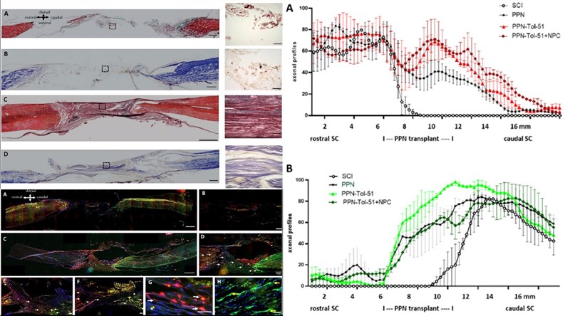 Fig. 2 The results of histological analysis, axonal tracing, and axonal regeneration.2
References
- Brocard, Frédéric, and Nejada Dingu. "Calpains at the Crossroads of Spinal Cord Physiology, Plasticity, and Pathology." Cells vol. 14,19 1503. Distributed under Open Access license CC BY 4.0, without modification. https://doi.org/10.3390/cells14191503
- Arriero-Cabañero, Alejandro et al. "Transplantation of Predegenerated Peripheral Nerves after Complete Spinal Cord Transection in Rats: Effect of Neural Precursor Cells and Pharmacological Treatment with the Sulfoglycolipid Tol-51." Cells vol. 13,16 1324. Distributed under Open Access license CC BY 4.0, with modification. https://doi.org/10.3390/cells13161324
For Research Use Only.