Leishmania derived Exosome Research & Application
Overview Advantages Research Application Services Products FAQs
Mechanistic Role of Exosomes in Leishmaniasis Pathogenesis
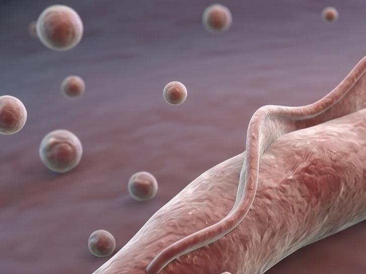
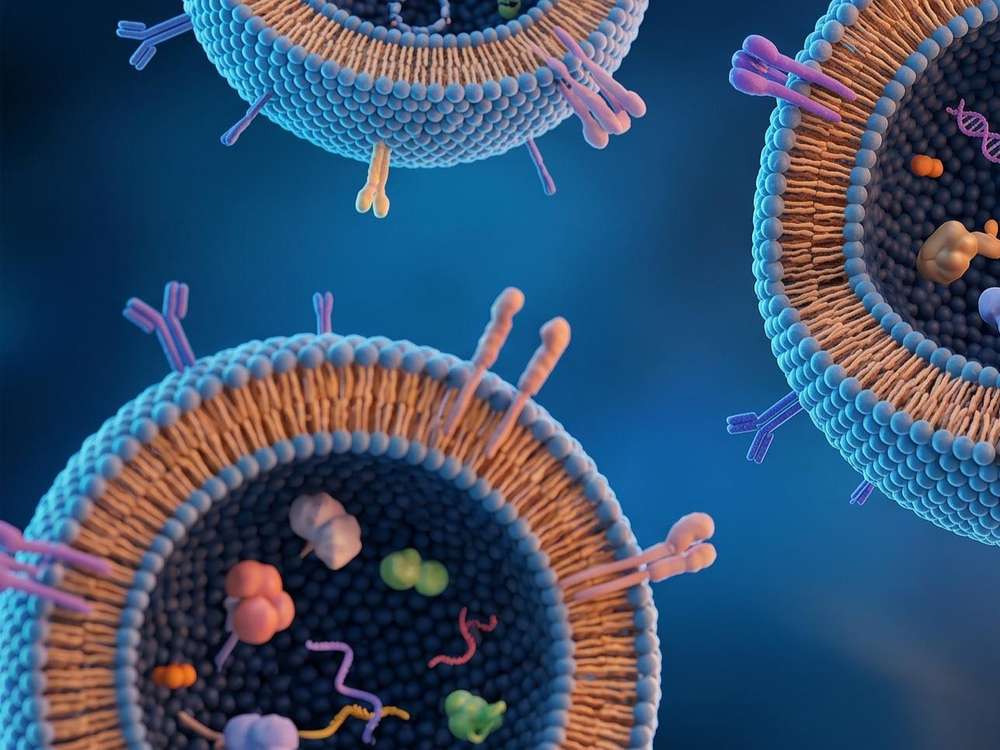
Leishmaniasis remains a significant global health burden, manifesting in clinical forms ranging from cutaneous lesions to fatal visceral systemic failure. The causative agent, a protozoan parasite of the genus Leishmania, has evolved sophisticated mechanisms to subvert host surveillance. Central to this biological evasion is the secretion of extracellular vesicles, specifically exosomes. These membrane structures act as critical conduits for intercellular communication, transporting a diverse cargo of proteins, small RNAs, and lipids from the parasite to host immune cells.
Deciphering Pathogenesis
Exosomes serve as primary vehicles for the delivery of virulence factors, such as the zinc metalloproteinase GP63, directly into the host cytosol.
Crosstalk between Host and Parasite
They provide insights into how eukaryotic pathogens manipulate mammalian signaling pathways without direct intercellular contact.
Dynamics in Vector and Host
Recent evidence confirms that these vesicles are swallowed with the parasite during the sand fly bite, making them an integral component of the initial infectious inoculum.
Therapeutic Innovation
Understanding the molecular architecture of these vesicles is the first step toward utilizing them as diagnostic biomarkers or scaffolds for vaccine delivery.
Consult With Us to Define Your Research Parameters
Distinctive Biochemical Properties of Parasite Derived Vesicles
Leishmania exosomes possess distinct biochemical and biophysical properties that distinguish them from typical mammalian vesicles, providing them with specialized functional advantages in the context of infection.
|
Characteristic
|
Biological Significance
|
|
Enzymatic Enrichment
|
High concentrations of GP63 and EF1α allow for the immediate deactivation of host tyrosine phosphatases upon entry.
|
|
Environmental Resilience
|
Stable within the harsh, alkaline environment of the sand fly midgut and the acidic phagolysosomes of macrophages.
|
|
Secretion Induced by Stress
|
Vesicle release is upregulated during temperature shifts (from 26°C to 37°C), mimicking entry into the mammalian host.
|
|
Selective Cargo Loading
|
Proteomic signatures differ between procyclic and metacyclic stages, indicating a highly regulated sorting mechanism.
|
|
Immune Evasion
|
Capable of traversing anatomical barriers to modulate distant dendritic cells and T lymphocytes before the parasite arrives.
|
Discuss Your Project Goals with Our Lead Exosome Researchers
Current Landscapes in Leishmania Exosome Research
The scientific community has made progress in characterizing the role of these vesicles across different species and lifecycle stages. Current literature underscores a dual role for Leishmania exosomes: they are both suppressors of specific microbicidal functions and activators of localized inflammation that favors the parasite.
Immune Modulation and Signaling Interference
Research indicates that vesicles from L. donovani and L. infantum selectively reprogram the cytokine profile of human monocytes. By promoting an antiphlogistic environment characterized by elevated IL10 and diminished TNFα, the vesicles ensure a permissive niche for amastigote replication. Concurrently, the delivery of effector proteins such as Annexin A1 facilitates parasitic internalization through the FPR1/2 receptor axis.
Proinflammatory Hyperpathogenesis
Counterintuitively, vesicles from L. major and L. braziliensis have been shown to exacerbate skin lesions by recruiting neutrophils and inducing proinflammatory mediators like IL17a and IL6. This localized inflammation, often mediated by the NLRP3 inflammasome, creates an environment that facilitates the spread of the pathogen rather than its clearance.
Viral Cargo and Gene Transfer
A critical niche in this research field involves the discovery of Leishmania RNA virus 1 (LRV1) within exosomes. These vesicles serve as protective carapaces for the virus, allowing it to trigger Toll like receptor 3 (TLR3) and drive a potent inflammatory response that correlates with the severe mucocutaneous form of the disease.
Partner With Our Scientists to Optimize Your Functional Analysis Study
Translation and Therapeutic Prospects
The translation of exosome research holds promise for shifting the management of Leishmaniasis from reactive treatment to proactive prevention and precision therapy.
 Vaccinology
Given their ability to carry native pathogen antigens in a highly immunogenic format, exosomes are being evaluated as vaccine candidates. They offer a safer alternative to attenuated strains while maintaining the structural integrity of complex epitopes.
Targeted Drug Delivery
The natural tropism of these vesicles for macrophages can be utilized to deliver drugs directly to the site of infection, reducing systemic toxicity and enhancing therapeutic efficacy.
Diagnostic Markers
The detection of specific RNA sequences or proteins within circulating exosomes could provide a noninvasive method for monitoring visceral Leishmaniasis progression or treatment response.
Neutralization Strategies
Developing small molecules or antibodies that block exosome biogenesis or uptake could represent a novel class of adjunctive therapies to prevent disease exacerbation.
Coordinate With Our Team to Tailor Your Exosome Research Strategy
Integrated Solutions for Protozoan Exosome Analysis
Creative Biolabs provides a specialized technical pipeline to support the global research community in navigating the complexities of vesicles derived from parasite. Our platform integrates advanced biophysical techniques with deep biological insights to provide solutions.

We utilize high resolution methodologies to ensure the purity and integrity of isolated vesicles:
Our laboratory is equipped for both in vitro and in vivo validation:
-
Macrophage Interaction Assays
Monitoring motility, chemotaxis, and phagocytic activity.
-
Cytokine Profiling
Multiplex analysis of the host secretome (IL10, IL12, TNFα, etc.) following vesicle exposure.
-
Signaling Pathway Analysis
Assessing the activation of host protein tyrosine phosphatases and the NLRP3 inflammasome.
We provide granular analysis of the exosomal cargo to identify potential virulence factors or biomarkers:
-
Proteomics
Comparative analysis of vesicle protein composition across different stages of the parasite lifecycle.
-
Transcriptomics
Identification of mRNA and miRNA derived from parasite that may influence host gene expression.
-
Lipidomics
Analysis of membrane lipids that facilitate fusion with host cells.
Connect With Our Experts to Accelerate Your Discovery Pipeline
High Performance Reagents for Exosome Investigation
In addition to our analytical services, Creative Biolabs offers specialized reagents designed to standardize exosome research. These products are manufactured under rigorous quality control to ensure reproducibility in scientific investigations.

Isolation Reagents
Optimized kits for the rapid recovery of vesicles from complex biological fluids or parasite conditioned media.

Labeling and Tracking Tools
Fluorescent dyes and specialized kits for monitoring vesicle internalization and intracellular trafficking within host cells.

Exosome Standards
High purity, characterized vesicle preparations to serve as positive controls in functional and analytical assays.
Speak With Us Regarding Your Requirements
FAQs
Q: What is the primary advantage of using exosomes over whole parasites in vaccine research?
A: Exosomes provide a focused antigenic profile and lack the infectious potential of whole parasites. They act as a sophisticated "antigen delivery system" that can be precisely characterized and standardized, reducing the variability often seen with whole organism preparations.
Q: Can I send you preisolated exosome samples for analysis?
A: Yes, we accept exosome samples isolated by customers. Upon receipt, our technical team will first perform a preliminary quality assessment to ensure that the samples meet the requirements for subsequent experiments, such as protein characterization or cell treatment assays, thereby ensuring the reliability and accuracy of the experimental results.
Q: Can you provide a basic report on exosome size and concentration?
A: Yes. This is one of our standard characterization services. Using Nanoparticle Tracking Analysis (NTA), we can provide a detailed report including the average particle size, particle size distribution profile, and the precise particle concentration per milliliter of sample.
Q: Can you help confirm whether exosomes contain a specific protein?
A: Yes. If you provide the name or sequence of the target protein, we can analyze your samples using mass spectrometry. We can assist in verifying whether the exosomes successfully carry a specific virulence factor or marker protein.
Q: After the experiment is completed, will I receive the original microscopy images and raw data files?
A: Yes. The final technical report will include all original experimental images, such as transmission electron microscopy (TEM) images showing exosome morphology, as well as all raw statistical data. These materials can be directly used for manuscript preparation, publication, or internal project reporting.
Ask Your Questions
For Research Use Only. Cannot be used by patients.
Related Services: