With the development of molecular imaging and material science, single domain antibody (sdAb)-targeted imaging technology has attracted extensive attention in the academic area. Many sdAb-based tracers using either nuclear, fluorescent labels or nanobubble/microbubble have been heavily investigated pre-clinically and are currently making their way into the clinic. Armed with rich experience in the production of sdAb and non-invasive molecular imaging research, Creative Biolabs provides customized nano-tracer development services with various labels. We provide tailored imaging tools to support your research in different fields ranging from cancer, inflammation with sdAbs, tumor extracellular matrix, to checkpoint molecules and immune responses.
Radiolabeled sdAbs for Molecular Imaging
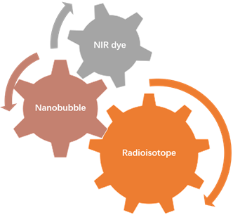
Nuclear molecular imaging requires the targeting moiety, in our case is a sdAb, to be labeled with a diagnostic radioisotope. The latter can either be a gamma-emitting isotope for single photon emission computed tomography (SPECT) or a positron-emitting isotope for positron emission tomography (PET). 99mTc-tricarbonyl reacts site-specifically with a genetically inserted C-terminal hexahistidine tag, which can also be used for purification purposes via immobilized metal affinity chromatography. Radiolabeling of proteins with other radiometals (such as 64Cu, 68Ga, and 89Zr for PET or 67Ga and 111In for SPECT) or radiohalogens (such as 18F and 124I for PET and 123/131I for SPECT) usually necessitates the use of a chelator for complexation or a prosthetic group for electrophilic substitution, respectively.
NIR dyes-conjugated SdAbs for Molecular Imaging
Different from radiolabeled imaging, fluorescence imaging requires the presence of a fluorescent label for sensitive detection. Though a wide range of fluorescent dyes are available for biotechnological purposes, for in vivo imaging applications, the choice of fluorophore is limited to those emitting in the near-infrared (NIR) region, and more specifically, those with a maximal excitation and emission wavelength between 650 and 900 nm. Commonly, hydrophilic (sulfonated) variants of cyanine dyes with a penta- or heptamethine chain are used.
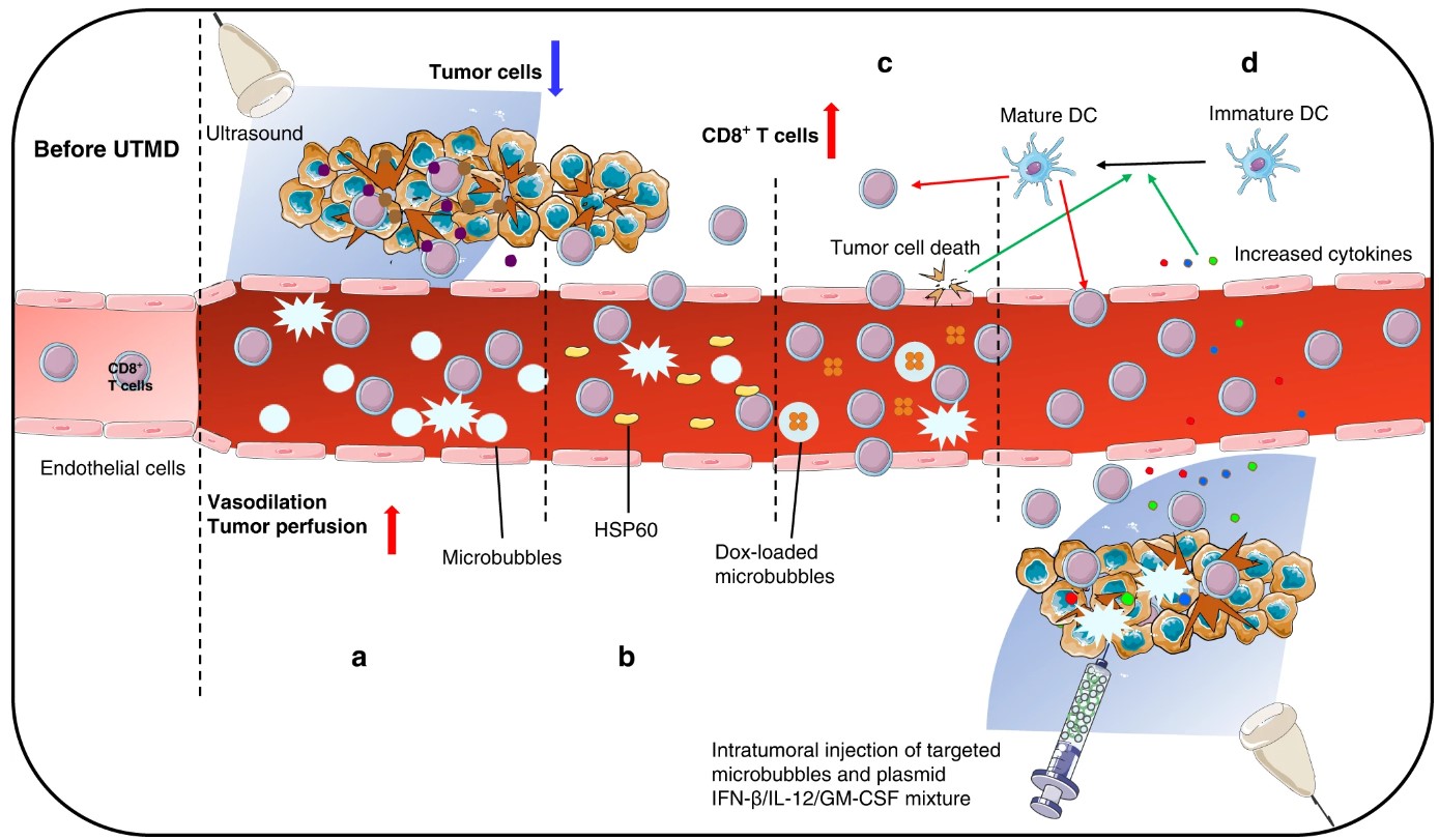 Fig.1 Enhancing immunotherapeutic efficacy through ultrasound-targeted microbubble destruction-mediated remodeling of the tumor microenvironment.1,4
Ultrasound-targeted Nanobubble for Molecular Imaging
Active targeting rapidly and selectively enhances tumor accumulation and tumor retention. These processes could be also visualized and quantified in real-time with ultrasound. Ultrasound imaging makes use of sound waves which are reflected differently by different tissues and organs. Microbubbles are generally used as contrasting agents and sdAb mediated guidance can further aid in improving contrast at the desired location. Due to the micrometer size, the proteins targeted should be located in the vasculature of the tumor such as VCAM-1 and PSMA. The application of sdAb-targeted microbubbles is beneficial since it can provide safe, fast and inexpensive scanning together with relatively high-quality images.
Services
- sdAb generation and optional modification
- Label design and conjugation
- Molecular imaging services in the disease model
- Comprehensive data analysis
If you are interested in our customized labeled nano-tracer development services, please directly contact us for more information.
Published Data
1. A Single-Domain Antibody for the Detection of Pathological Tau Protein
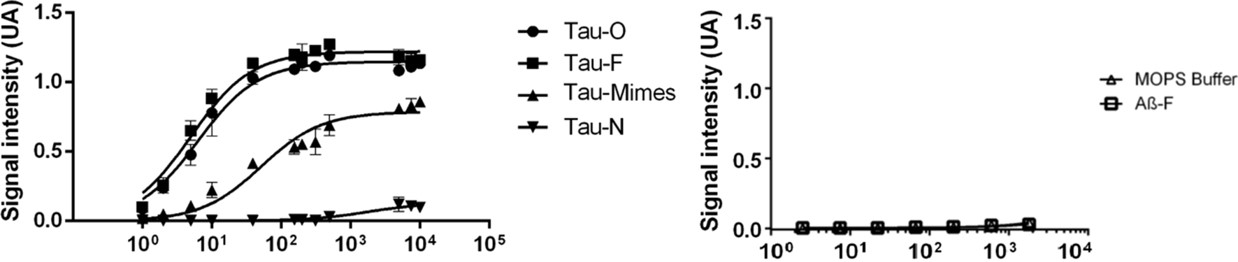 Fig.2 ELISA assays for anti-Tau sdAb with different species.2
This study introduced a sdAb as a novel radiotracer for detecting and monitoring oligomeric Tau species in the human brain. The anti-Tau sdAb showed strong binding to Tau oligomers (Kd = 6.280 nM) and Tau fibers (Kd = 5.024 nM), with weaker binding to native Tau (Kd = 1791 nM) and amyloid peptide (Kd > 10,000 nM). The sdAb facilitated high-contrast immunohistological labeling of Tau pathology in neurons and plaques in Alzheimer’s disease (AD) patients. When radiolabeled with 99mTc, the sdAb maintained stability (radiochemical purity > 93%) for up to 6 hours. However, in healthy mice, most of the injected dose was taken up by the kidneys, with minimal brain uptake (0.17% ID/g at 5 min). Despite this, the sdAb holds promise for preclinical imaging of Tau oligomers, which may serve as an early marker of AD pathology.
2. Noninvasive In Vivo Imaging of α-Synuclein or Tau Pathology via Single-Domain Antibody
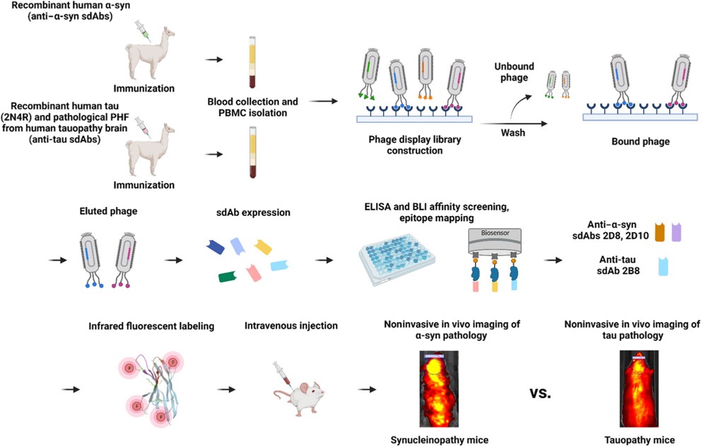 Fig.3 A summary of developing the anti–α-syn and anti-tau sdAb imaging probes.3,4
In this study, researchers developed sdAb-based imaging probes that specifically target α-synuclein (α-syn) and tau lesions, enabling their in vivo detection in animals. Two anti–α-syn sdAbs demonstrated strong brain signals in M83 α-syn mice after peripheral injection, with no signal in tauopathy or WT mice. These signals correlated closely with insoluble α-syn levels postmortem (syn 211: r = 0.958 and 0.927; PA5-13401: r = 0.978 and 0.955, P < 0.0001). Similarly, the anti-tau sdAb provided strong brain signals in tauopathy models, correlating with both insoluble (CP27: r = 0.954, P < 0.0001) and soluble tau levels (CP27: r = 0.932, P < 0.0001; PHF1: r = 0.733, P = 0.0001). Besides, both sdAbs colocalized specifically with their respective target proteins, α-syn and tau, in the brain.
References
- Liu, Senbo, et al. "Ultrasound-targeted microbubble destruction remodels tumour microenvironment to improve immunotherapeutic effect." British Journal of Cancer 128.5 (2023): 715-725.
- De Leiris, Nicolas, et al. "A single-domain antibody for the detection of pathological Tau protein in the early stages of oligomerization." Journal of Translational Medicine 22.1 (2024): 163. Distributed under Open Access license CC BY 4.0. The image was modified by extracting and using only part of the original image.
- Jiang, Yixiang, et al. "Single-domain antibody–based noninvasive in vivo imaging of α-synuclein or tau pathology." Science advances 9.19 (2023): eadf3775.
- Distributed under Open Access license CC BY 4.0, without modification.
For Research Use Only.