Creative Biolabs is a world-leading service provider of application-specific antibody development. Especially, we offer high-quality in vitro diagnostic (IVD) antibody and immunoassay development services targeting a wide range of disease biomarkers. Here, we focus on the NfL as a biomarker of neuronal injury.
Introduction to NfL
Neurofilaments (Nfs) are neuron-specific cytoskeletal proteins with a characteristic diameter of 8-10 nm. They are present in cell bodies, dendrites, and axons and are involved in the radial growth, stabilization, and polarization of neural cells, enabling effective axonal conduction. According to the molecular mass of their subunits, neurofilaments are classified as light (NfL), middle (NfM), heavy chain (NfH), and α-internexin (α-int). All of these subunits have a conserved α-helical rod domain with a variable amino-terminal and carboxy-terminal region. The length of the tail confers a different molecular weight. The nomenclature-light (~68 kDa), -medium (~145 kDa), and heavy (~200 kDa) refer to the molecular weight of the filaments. Among the three subunits, NfL is the backbone of Nfs, representing the most abundant subunit and the most soluble one.
Under normal conditions, low levels of NfL are constantly released from axons to biological fluids, probably in an age-dependent manner, with higher levels of NfL being released at older ages. However, following axonal damage in the central nervous system (CNS) that are caused by different diseases, the release of NfL sharply increases. The level increases proportionally in the cerebrospinal fluid (CSF) and blood to the degree of axonal damage both in normal and in pathologic conditions. The pathological conditions include a variety of neurological disorders, such as inflammatory, neurodegenerative, traumatic, and cerebrovascular diseases.
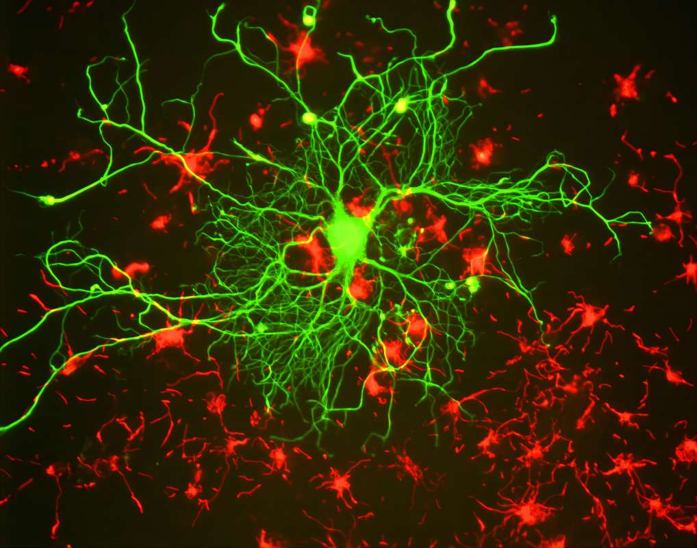 Fig.1 Immunostaining of cortical neuron with NF-L antibody in green.1
NfL as A Diagnostic Marker of Neurological Diseases
The value of NfL as a diagnostic, prognostic, and therapeutic monitoring biomarker in neurological diseases has been highlighted by different studies. The role of NfL as a biomarker has been largely reported in multiple sclerosis (MS), Alzheimer's disease (AD), frontotemporal dementia (FTD), amyotrophic lateral sclerosis (ALS), atypical parkinsonian disorders (APD), Creutzfeldt-Jakob disease (CJD), Hiv-associated dementia (HAD), and traumatic brain injury (TBI).
Both CSF and blood levels of NfL indicate disease severity and/or progression. Detection of NfL in CSF can be achieved by using sandwich ELISA technology or immunoblot. Due to the fact that the concentration of NfL in the blood is roughly 40-fold lower than that in the CSF, the sensitivity of ELISA is not sufficient for blood NfL measurement. New immunoassays, therefore, have been developed to enable biomarker detection at ultralow levels and allow for the measurement of NfL in blood, such as electrochemiluminescence (ECL) assay. This makes it possible to easily and repeatedly measure NfL for monitoring diseases' courses.
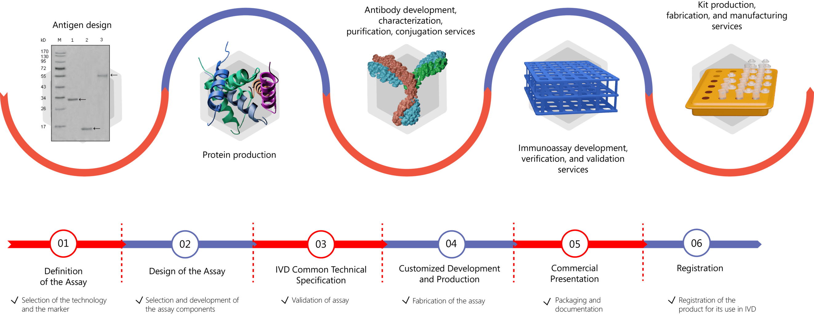
IVD Antibody/Immunoassay Development Services Provided by Creative Biolabs
As NfL represents one of the most promising biomarkers in different neurological disorders, appropriate antibodies and sensitive immunoassays can be generated to improve the diagnostic accuracy and prognostic assessment, as well as management. The detection and quantification of biomarkers largely rely on the use of sensitive immunoassays, the core elements of which are antibodies. Through our role as a leading antibody service provider, Creative Biolabs is specialized in the development of biomarker-specific antibodies with high quality. Besides, we also offer immunoassay development services of various immunoassay formats, such as ELISA, ECL, immuno-PCR, and lateral flow assays. Please click the links below for more information:
- IVD Antibody Development
- Antibody Pair Development
- Antibody & Protein Conjugation
- IVD Immunoassay Development
Features of Our Services
- Flexibility in antibody/immunoassay development services to suit the specific needs of the clients
- Abundant expertise with know-how in all steps of the research, development, verification, and validation process
- Quick response and expert technical support from inquiry to project completion
- Cutting-edge technologies, in-depth expertise, and reasonable prices
Please feel free to contact us for more information and discuss your project requirements.
Reference
- From Wiki: By GerryShaw - Own work, CC BY-SA 3.0, https://commons.wikimedia.org/wiki/File:Neuron_in_tissue_culture.jpg
For Research Use Only.