Surgically induced Osteoarthritis (OA) Model
Creative Biolabs offers a battery of rodent models for disease mechanism studies as well as the screening of therapeutic candidates. We have talented experts and scientists who are willing to help to set up a detailed research plan based on your specific needs. Herein, you can find the most comprehensive services with the most reasonable prices.
Introduction of Osteoarthritis
Osteoarthritis (OA) is a joint disease that involves degeneration of cartilage, limited synovitis, subchondral bone changes, and osteophyte formation. According to the disease etiology, OA has been divided into two categories: primary OA (POA) and secondary OA. POA is a naturally occurring phenomenon due to degenerative changes in the joint, while the latter a condition occurring in the presence of specific causes or risk factors, including trauma, congenital diseases, and other diseases or disorders of metabolism or the bone. Post-traumatic osteoarthritis (PTOA), as the most widely studied subcategory of secondary OA, results from an injury to the affected joint.
Animal models of OA can be broadly divided into spontaneous and induced models. Spontaneously OA models involve naturally occurring and genetic models of disease, while induced models are further classified into surgically induced and chemically induced models. Each model simulates different aspects of human OA disease, playing a key role in understanding its pathophysiology as well as developing successful treatment regimens.
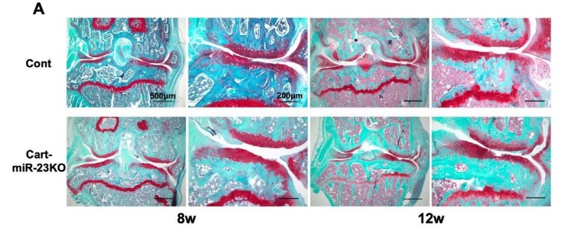 Fig. 1 Safranin-O staining in the knee joints of Control and Cart-miR-23clus KO mice in surgically induced OA model.1
Surgically-Induced OA Models
Surgical methods of OA induction work through a combination of joint destabilization, altered joint mechanics, and intra-articular inflammation. These methods create models that are considered to be more appropriate to represent POA in humans. Commonly used surgically-induced OA models include anterior cruciate ligament transection (ACLT), partial or total meniscectomy, medial meniscal tear (MMT), and ovariectomy. These models progress rapidly, making them excellent choices for short-term studies. Further, these models have the advantage of reproducibility compared to spontaneous models.
Anterior Cruciate Ligament Transection (ACLT) Rat Model
ACLT model is induced due to ACL injury, which causes joint destabilization, subsequently leading to PTOA. Technically, the skin is prepped with a topical antiseptic following anesthesia, and an incision is made in the skin laterally to the right knee joint. After the joint capsules are opened, the ACL is transected using a surgical scalpel. The anterior drawer test is used to test the success of this procedure and the left knee joint is the ACL-intact control joint. This model is suitable for evaluating bone resorption inhibitors as potential disease-modifying pharmaco-therapies.
Medial Meniscal Tear (MMT) Rat Model
The MMT model in animals is achieved through transection of the medial collateral ligament in the knee. The MMT-induced joint damage model in rats has many features attractive for an animal model:
- These animals show joint instability and tibial cartilage damage in as little as 7-14 days after surgery.
- MMT-induced joint damage lesions include articular cartilage proteoglycan loss, chondrocyte degeneration and loss of matrix.
- This model is useful to evaluate potential therapeutic agents.
In testing the therapeutic efficacy of drug candidates, we are capable of conducting all kinds of assessments including but not limited to:
- Gait analysis
- Clinical assessment
- Histopathological assessment
- Immunohistochemistry analysis
- Cytokine/Chemokine analysis
- Micro-CT
- Pain assessment
Additionally, other examples of rodent musculoskeletal disease models that you may be interested include:
- Monosodium Iodoacetate (MIA)-Induced Osteoarthritis Model
- Fracture Models
- Bone Defect Models
- Orchiectomy-Induced Osteoporosis Model
- Ovariectomy (OVX)-Induced Osteoporosis in Rats
- Adjuvant-Induced Arthritis (AIA) Rodent Model
- Collagen-Induced Arthritis (CIA) Rodent Model
- Collagen Antibody-Induced Arthritis (CAIA) Model
Creative Biolabs offers turn-key or ala carte services customized to our client's every specific requirement. Please feel free to contact us or send us an inquiry, if you are interested in any of these services.
Reference
- Fujiwara, Yusuke, et al. "miR-23a/b clusters are not essential for the pathogenesis of osteoarthritis in mouse aging and post-traumatic models." Frontiers in Cell and Developmental Biology 10 (2023): 1043259. doi:10.3389/fcell.2022.1043259. Distributed under Open Access license CC BY 4.0. The image was modified by extracting and using A part of the original image.
For Research Use Only.