x
-
Research Models
-
Ex Vivo Tissue Model
- Ex Vivo Human Genitourinary Tissue Model
- Ex Vivo Human Endocrine System Tissue Model
- Ex Vivo Human Cardiovascular Tissue Model
- Ex Vivo Human Skin System Tissue Model
- Ex Vivo Neurological System Model
- Ex Vivo Human Digestive System Model
- Ex Vivo Human Musculoskeletal System Tissue Model
- Ex Vivo Human Respiratory System Tissue Model
- Precision-Cut Tissue Slicing Model
- 3D Spheroid Model
- 3D Organoid Model
- Organ-on-a-Chip Model Introduction
-
Ex Vivo Tissue Model
-
Services
- 3D Biology Based Biomarker Discovery Services
-
3D Biology Based Drug Discovery and Development Services
- 3D Biology Based Immuno-Oncology Drug Discovery and Development
- 3D Biology Based Neurological Disorder Drug Discovery and Development
- 3D Biology Based Inflammation & Immunological Disease Drug Discovery and Development
- 3D Biology Based Metabolic Disease Drug Discovery and Development
- 3D Biology Based Urological System Disease Drug Discovery and Development
- 3D Biology Based Digestive System Disease Drug Discovery and Development
- 3D Biology Based Ocular Disease Drug Discovery and Development
- 3D Biology Based Musculoskeletal Disease Drug Discovery and Development
-
3D Biology Based Toxicity Evaluation Services
- 3D Biology Based Ocular Toxicity Evaluation Services
- 3D Biology Based Skin Toxicity Evaluation Services
- 3D Biology Based Cardiotoxicity Evaluation Services
- 3D Biology Based Hepatotoxicity Evaluation Services
- 3D Biology-Based Neurotoxicity Evaluation Services
- 3D Biology-Based Nephrotoxicity Evaluation Services
- 3D Biology-Based Gastrointestinal Toxicity Evaluation Services
- 3D Biology Based Phototoxicity & Photoallergy Evaluation Services
- 3D Biology Based Pancreatic Endocrine Toxicity Evaluation Service
- 3D Biology Based Inhalation Toxicity Evaluation Services
- 3D Biology Based Oral Irritation Evaluation Services
- Supporting Assay Services
-
Products
- Body Fluids
-
Cells
- Cardiomyocytes
- Bone Cells & Chondrocytes
- Neural Cells
- Preadipocytes & Adipocytes
- Peripheral Blood Mononuclear Cells (PBMCs)
- Purified Immune Cell Populations
- Tissue Resident Immune Cells
- Bone Marrow-derived Cells
- Primary Epithelial Cells
- Primary Endothelial Cells
- Primary Fibroblasts
- Primary Smooth Muscle Cells
- Macrophages
- Stem Cells
- Hepatocytes and Non-Parenchymal Cells
- Islet Cells
- GFP-expressing Stable Cells
- RFP-expressing Stable Cells
- Luciferase-expressing Stable Cells
- Cas9-expressing Stable Cells
- Cre-expressing Stable Cells
- Immortalized GFP-expressing Stable Cells
- Immortalized Primary Cells
- Genetically Modified Cells
- 3D Models
- 3D Cultures
- Nucleic Acids
- Subcellular Samples
- Tissues
- Cell & Tissue Lysates
- Recombinant Proteins
- Resource
- Company
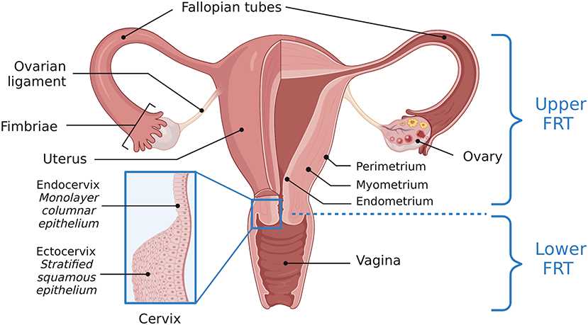 Fig 1. Anatomy of the female reproductive tract.1,3
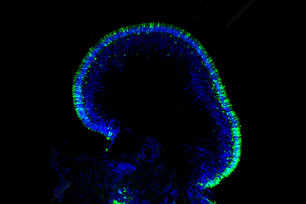
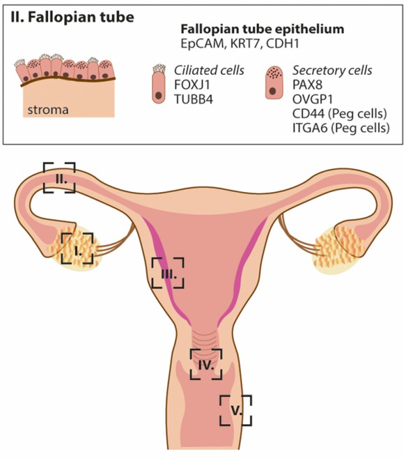 Fig 2. The main cell types of the fallopian tube and their typical markers.2,4
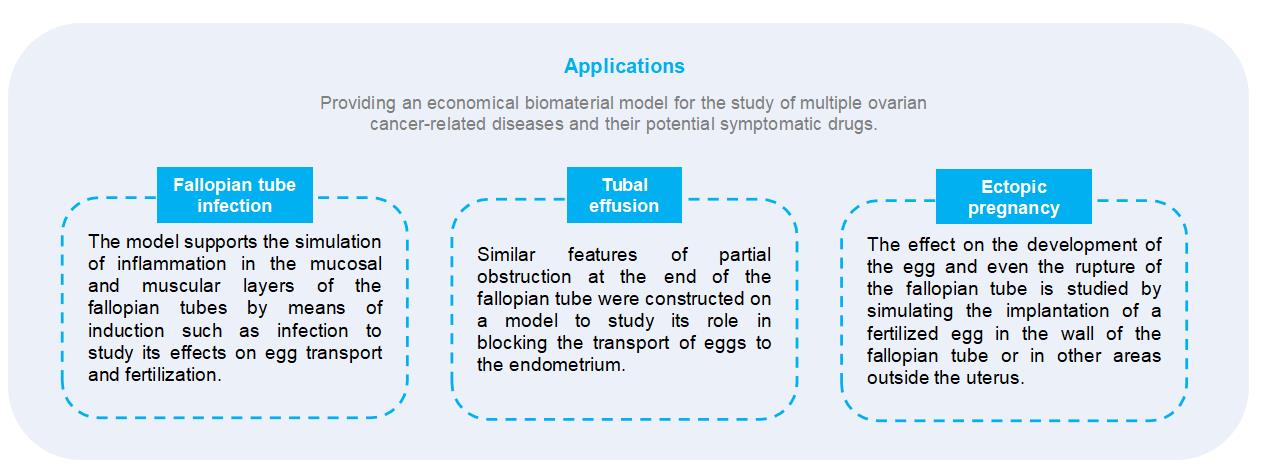
 Fig 3. The applications of fallopian tube organoid models.2,3