
Mechanism Discovery
Profile pathway activation and map complement signals to osteogenic, osteoclastogenic, inflammatory, and repair responses.


Creative Biolabs provides integrated research services to define how complement drives bone loss, remodeling, repair, and osteoimmune signaling, and to evaluate complement-targeted therapeutics in osteoporosis, fracture healing, inflammatory osteolysis, arthritis-associated bone damage, and periodontal bone disease. Support spans target and biomarker selection, complement profiling, osteoblast/osteoclast assays, bone-relevant cell and animal models, mechanism-of-action studies, candidate efficacy testing, and data interpretation.
Discuss Your Osteology Research PlanCreative Biolabs supports studies in which complement activation is suspected to influence osteoclast formation, osteoblast signaling, bone resorption, mineralization, fracture repair, or inflammatory damage at the bone–immune interface. We help researchers move from a broad disease hypothesis to a measurable study plan by selecting the relevant complement pathway, bone-cell system, activation matrix, disease stimulus, pharmacologic comparator, and bone outcome.
Projects are suitable for early target discovery, translational biomarker work, lead selection, mechanism-of-action confirmation, and preclinical efficacy evaluation. A study may begin with patient serum or plasma, an existing antibody or inhibitor, a proposed target such as C3aR or C5aR1, or a bone-disease model that needs complement-resolved endpoints. Complement testing can be combined with osteoblast, osteoclast, stromal-cell, immune-cell, tissue, and in vivo readouts in one coordinated program.
Profile pathway activation and map complement signals to osteogenic, osteoclastogenic, inflammatory, and repair responses.
Interrogate components, receptors, regulators, or pathway nodes using inhibition, depletion, stimulation, or genetic strategies.
Rank antibodies, proteins, peptides, aptamers, or small-molecule inhibitors in orthogonal functional assays.
Combine complement biomarkers with bone turnover, cell phenotype, imaging, histology, and biomechanical endpoints.
We can translate the disease biology, available samples, and intended therapeutic claim into a staged complement–bone research strategy.
Complement can be protective during early repair yet damaging when activation is excessive, persistent, mistimed, or localized to vulnerable bone and joint surfaces. We therefore define the disease phase and cellular source of the phenotype before choosing assays or therapeutic endpoints.
Investigate complement-dependent changes in RANKL/OPG balance, osteoclastogenesis, osteoblast signaling, trabecular structure, bone turnover markers, and inflammatory mediators in estrogen-deficiency or aging-related settings.
Review the C5aR Therapeutic TargetResolve complement activity across inflammatory, repair, and remodeling phases; evaluate callus formation, mineralization, osteoclast activity, immune recruitment, and mechanical recovery; and compare normal healing with delayed union or non-union biology.
Explore Complement Activation Product TestsStudy complement-mediated osteoclast activation, cytokine amplification, cartilage–bone interface injury, immune-complex activation, and candidate response in arthritis or other inflammatory osteolysis contexts.
Explore Rheumatoid Arthritis ResearchAssess dysregulated complement signaling in bacteria-driven inflammation, connective-tissue disease, alveolar bone resorption, and osteoclastogenic responses using serum, gingival, immune-cell, or bone-cell models.
View Periodontal EDS Research SupportDetermine whether a material surface initiates complement deposition, anaphylatoxin release, inflammatory-cell recruitment, or altered osteoblast/osteoclast behavior, and test coating or inhibitor strategies.
View C3b Deposition AssaysCompare variants, patient-derived matrices, complement deficiencies, or regulator abnormalities when the mechanism is uncertain. Feasibility work can establish whether the phenotype is pathway-, receptor-, or cell-context dependent.
Explore Complement Genetic TestingA pathway assay confirms that complement changed; a bone-cell assay shows why that change matters. Modules can be used independently or assembled into a mechanism-led package with shared controls, matrices, concentrations, and time points.
| Research module | Representative capabilities | Decision supported |
|---|---|---|
| Complement pathway profiling | Classical, alternative, and lectin pathway activity; CH50/AP50-style functional testing; pathway-selective activation or inhibition | Identify the active pathway, baseline activity, sample suitability, and degree of pathway suppression |
| Activation products and deposition | C3a, C5a, Bb, C4d, sC5b-9, C3b/iC3b, C4b, and C5b-9 measurements in serum, plasma, supernatant, cells, tissue, or material surfaces | Locate the cascade step affected by disease or treatment and distinguish soluble activation from surface injury |
| Osteoclast formation and resorption | Precursor differentiation, TRAP staining/activity, multinucleation, actin-ring formation, NFATc1/cathepsin K markers, resorption-pit assays, and RANKL/OPG response | Determine whether complement or a candidate changes osteoclast commitment, maturation, or functional bone resorption |
| Osteoblast and stromal responses | Viability, proliferation, migration, ALP activity, mineralization, RUNX2/osteocalcin expression, cytokines, chemokines, RANKL/OPG, and receptor expression | Define direct effects on bone-forming cells and indirect control of osteoclastogenesis |
| Osteoimmune co-culture | Osteoblast–osteoclast, MSC–myeloid, macrophage–bone cell, or client-defined co-cultures with complement-active or controlled matrices | Capture cell–cell feedback that is missed in isolated target-binding or single-cell assays |
| Tissue and structural endpoints | Histology, immunohistochemistry, TRAP staining, micro-CT, bone morphometry, serum bone-turnover markers, and biomechanical testing where applicable | Translate molecular and cellular effects into bone architecture, repair quality, and functional strength |
Multiplex and orthogonal single-analyte testing can profile dose response, time course, pathway engagement, and sample-to-sample variation.
Model choice follows the decision point. Purified systems clarify direct complement mechanisms; cell systems reveal osteoimmune signaling; tissues and in vivo models establish spatial, temporal, structural, and functional relevance.
Profile complement activation and bone-related biomarkers in serum, plasma, synovial fluid, conditioned medium, or project-defined samples while controlling collection, anticoagulant, freeze–thaw, storage, and ex vivo activation variables.
Use primary cells, established cell lines, mesenchymal stromal cells, or client-provided cells to evaluate receptor expression, differentiation, mineralization, migration, inflammatory signaling, and osteoclast-supporting factors.
Differentiate monocyte or marrow-derived precursors under defined osteoclastogenic conditions, introduce complement ligands or candidate therapeutics, and quantify both cell formation and mature resorptive function.
Combine bone cells with macrophages, monocytes, neutrophils, or other immune populations to evaluate complement-dependent recruitment, cytokine circuits, cell-contact effects, and bidirectional coupling.
Evaluate complement deposition and bone-cell response on biomaterials, extracellular matrix, bone slices, tissue sections, or three-dimensional constructs where surface chemistry and spatial context influence activation.
Tailor studies to osteoporosis, inflammatory bone loss, fracture repair, periodontal bone loss, or a client-defined indication, with complement pharmacodynamics linked to bone structure and function.
Candidate testing is structured so that loss of a bone phenotype can be attributed to a defined complement mechanism rather than nonspecific cytotoxicity, depleted serum activity, or assay interference.
Confirm identity, binding, competition, receptor occupancy, or component inhibition using ELISA, SPR/BLI, ligand–receptor assays, or project-specific biochemical formats.
Measure pathway inhibition, activation-fragment suppression, convertase or deposition effects, hemolysis, or cell-surface protection at matched candidate concentrations.
Determine whether the candidate changes osteoclast formation/resorption, osteoblast signaling/mineralization, RANKL/OPG balance, or osteoimmune cytokine networks.
Link pharmacodynamic complement markers to micro-CT, histology, bone turnover, callus composition, or mechanical outcomes in the relevant disease model.
Controls may include isotype or vehicle, inactive analog, heat-inactivated serum, complement-depleted and reconstituted serum, receptor agonist/antagonist, pathway-selective positive control, and cell-health controls. Where inhibition is potent, residual host-defense activity can be assessed with pathway-specific functional testing.
Explore Complement Inhibitor ValidationNeed study reagents? Browse complement antibodies, complement proteins, complement inhibitors, and sera and plasma products.
C3a, C5a, Bb, C4d, sC5b-9, pathway activity, component consumption, and tissue or surface deposition.
PINP/P1NP, osteocalcin, ALP, CTX-I, TRAP5b, RANKL, OPG, osteoblast/osteoclast numbers, and resorption activity.
IL-6, TNF, CXCL chemokines, immune infiltration, mineralization, bone volume, trabecular measures, callus composition, and strength.
Our complement-targeting development and testing capabilities can be combined into a single milestone-based program.
The workflow is adapted to the starting material and development stage. Early checkpoints reduce unnecessary animal work and ensure that advanced models use an active candidate, a responsive complement matrix, and validated bone endpoints.
Specify indication, disease phase, target cell, complement node, proposed mechanism, available sample or candidate, comparator, and success criteria.
Qualify matrix, complement activity, disease stimulus, bone-cell response range, controls, sampling time, and analytical readouts.
Generate concentration–response and time-course data across complement and bone-cell endpoints; test pathway or receptor dependence.
Transfer the selected condition into co-culture, tissue, biomaterial, or in vivo models with pharmacodynamic and structural endpoints.
Deliver raw data, quality control, statistical analysis, figures, interpretation, candidate ranking, and recommendations for the next study.
Useful starting information includes the disease indication and stage, proposed complement target, sample species and matrix, cell or model preference, candidate format and concentration range, expected route or timing of intervention, available benchmark, required bone outcomes, and the decision the dataset must support.
If the complement target is unknown, a discovery package can begin with pathway and activation-product profiling before committing to a candidate-specific efficacy study.
These studies illustrate how complement receptor activity and systemic pathway profiles can be connected to osteoclastogenesis, osteoporotic bone loss, fracture repair, and non-union biomarkers.
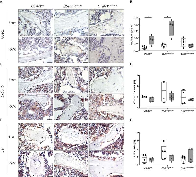
Bülow and colleagues used bone-cell-specific C5aR1 deletion and an ovariectomy model to show that osteoblast C5aR1 was required for osteoporosis-associated RANKL elevation and bone resorption, supporting cell-specific complement target validation.
View research via DOI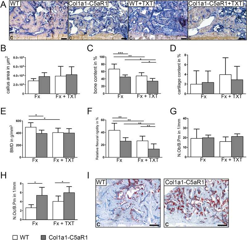
Bergdolt and colleagues connected C5aR1 signaling to reduced bone content, mineral density, and flexural rigidity with increased osteoclast numbers, illustrating an integrated cellular, histologic, imaging, and biomechanical efficacy framework.
View research via DOI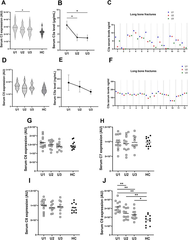
El-Sherbiny and colleagues profiled human long-bone fracture samples across inflammation, repair, and remodeling, identifying time-dependent complement changes and elevated MASP1 in non-union as a translational biomarker and target hypothesis.
View research via DOIWe support complement-focused studies in osteoporosis, fracture healing and non-union, inflammatory bone and joint damage, osteolysis, periodontal and craniofacial bone disease, bone–implant interactions, and project-defined rare or genetic bone phenotypes. Programs can focus on mechanism discovery, target validation, biomarker profiling, therapeutic candidate testing, or preclinical efficacy.
Yes. A discovery phase can compare classical, lectin, alternative, amplification-loop, and terminal-pathway activity using functional assays, activation products, deposition measurements, and disease-relevant samples. Results can then guide target selection and the design of bone-cell or efficacy studies.
Options include osteoclast differentiation, TRAP activity, actin-ring and resorption-pit assays; osteoblast viability, migration, ALP activity and mineralization; RANKL/OPG and cytokine analysis; and osteoblast–osteoclast or osteoimmune co-cultures. The exact panel is chosen according to the proposed mechanism and disease stage.
Yes. Candidate studies can include target engagement, receptor-dependent signaling, pathway pharmacodynamics, osteoclast and osteoblast responses, and advanced osteoporosis or fracture-healing endpoints. Controls are incorporated to distinguish target-specific activity from cytotoxicity, matrix effects, or global complement depletion.
Yes, subject to sample suitability and availability. Study design considers anticoagulant, collection and processing time, storage, freeze–thaw history, ex vivo complement activation, matrix interference, and the amount needed for repeat and orthogonal testing. Feasibility testing is recommended before a large cohort is analyzed.
Complement markers such as C3a, C5a, Bb, C4d, sC5b-9, and pathway activity can be analyzed alongside RANKL/OPG, CTX-I, TRAP5b, PINP, osteocalcin, cytokines, cell differentiation and resorption, histology, micro-CT, callus composition, or biomechanics. Shared time points and matched samples enable integrated interpretation.
Please provide the indication and disease stage, proposed target or hypothesis, species and sample matrix, available cell or animal model, candidate format and amount, preferred dose range, comparator, required endpoints, timeline, and the decision the study must support. If some items are unknown, they can be resolved during a feasibility and study-design phase.
Complement Activity/Function Assay Products
Learn More
Complement Testing Services
Learn More
Complement Therapeutics Featured Products
Learn More
Complement Therapeutics Services Brochure
Learn More
Aptamer Development PLATFORM
Learn More
ComPLETTM Hemolysis Assay Solutions
Learn More