Nephropathy & Complement Therapeutic Research Introduction

Nephropathy & Complement Therapeutic Research

Creative Biolabs provides complement-focused nephropathy research services for programs investigating immune-complex glomerular injury, alternative-pathway dysregulation, thrombotic microangiopathy, podocyte and tubular damage, renal complement deposition, genotype or autoantibody-driven disease, translational biomarkers, and complement-targeted therapeutics. We support experimental planning, sample and pathway qualification, renal cell and tissue models, mechanism-of-action studies, candidate testing, and integrated data interpretation.

Discuss Your Nephropathy Research Program
Glomerular injuryAlternative pathwayRenal biomarkersCandidate efficacy
Overview Renal Complement Disease Programs Assay Platform Models & Samples Candidate Evaluation Related Research FAQs Download Inquiry
Nephropathy Research Services

Build a Kidney-Relevant Complement Study around the Decision You Need to Make

Our services are designed for nephrology programs that need to determine which complement pathway is active, whether dysregulation is systemic or kidney-localized, how a genetic variant or autoantibody changes regulation, and whether a therapeutic candidate produces pathway-selective renal protection.

What we provide

Classical, lectin, alternative, and terminal-pathway profiling; activation-product and deposition assays; functional genetics; complement autoantibody studies; renal cell and tissue models; biomarker panels; and candidate efficacy evaluation.

Research needs supported

C3 glomerulopathy, immune-complex glomerulonephritis, IgA nephropathy, lupus nephritis, membranous nephropathy, complement-mediated HUS/TMA, FSGS, transplant-associated injury, and project-defined renal disease models.

Support delivered

Feasibility assessment, control and matrix planning, assay qualification, study execution, raw and processed data, statistics, images, mechanistic interpretation, biomarker or candidate ranking, and next-step recommendations.

Have a renal sample set but no qualified complement panel?

We can begin with pre-analytical risk review, matrix testing, pathway screening, and a staged biomarker plan.

Request Nephropathy Study Design Support
Mechanistic Framework

Resolve Complement Initiation, Amplification, Deposition, and Renal Injury

The same plasma marker can reflect different renal mechanisms. Our study design connects soluble pathway activity to the kidney compartment where complement is deposited, regulated, or converted into inflammatory and cytotoxic injury.

01

Trigger

Immune complexes, aberrant IgA, damaged endothelium, exposed matrix, ischemia, glycated surfaces, infection-related signals, or transplant-associated injury initiate complement.

02

Amplification

C3 convertase formation, factor B and factor D activity, properdin stabilization, nephritic factors, or insufficient factor H and factor I regulation sustain pathway output.

03

Renal deposition

C3 fragments, C4d, immunoglobulin-associated complement, and C5b-9 accumulate in glomerular, tubular, interstitial, or vascular compartments.

04

Tissue response

C3a/C5a signaling, endothelial activation, podocyte stress, mesangial inflammation, tubular injury, thrombosis, proteinuria, sclerosis, or fibrosis define the phenotype.

Mechanistic questions

Is low C3 caused by consumption? Does a patient sample stabilize C3 or C5 convertase? Is C5b-9 generated systemically or on renal cells? Does factor H-related protein competition reduce surface regulation? Which pharmacodynamic marker changes before the renal phenotype?

Causality controls

Complement-depleted and reconstituted serum, heat-inactivated matrix, pathway-selective buffers, wild-type and variant regulators, autoantibody-positive and negative samples, target-specific inhibitors, inactive or isotype controls, and rescue experiments.

Renal Research Contexts

Configure the Study for the Nephropathy Mechanism

Disease labels alone do not define the assay. We align pathway emphasis, sample matrix, renal compartment, controls, and efficacy endpoints with the proposed driver and disease stage.

C3 Glomerulopathy

Profile alternative-pathway dysregulation, C3 nephritic factors, factor H and FHR balance, genetic variants, convertase stability, C3 fragment deposition, and terminal-pathway activity.

Explore C3 glomerulopathy research

Complement-Mediated HUS and TMA

Study endothelial complement activation, regulatory variants or antibodies, C5a and C5b-9 formation, thrombosis-related endpoints, and rescue by proximal or terminal inhibitors.

Explore HUS complement research

Immune-Complex Glomerulonephritis

Assess classical, lectin, and alternative pathway contributions in IgA nephropathy, lupus nephritis, membranous nephropathy, and project-defined immune-complex models.

Explore glomerulonephritis research

Podocyte and Sclerotic Injury

Connect IgM/C3 deposition, soluble activation products, podocyte stress, permeability, proteinuria-related conditions, and sclerosis or fibrosis endpoints in FSGS-related research.

View C3b deposition assays

Tubular, Ischemic, and Metabolic Injury

Investigate complement activation on tubular epithelial cells or matrix under hypoxic, oxidative, glycated, toxic, or protein-overload stress and quantify inflammatory or viability outcomes.

Explore cell-based complement evaluation

Transplant-Associated Renal Injury

Evaluate donor-specific or project-defined antibody-mediated complement activation, endothelial deposition, C4d and C5b-9, inflammatory signaling, and inhibitor response.

View complement inhibition assays
Integrated Assay Platform

Measure Complement Function, Renal Deposition, and Phenotype with Matched Controls

Assay modules can be commissioned independently or combined into a staged study. Conditions are harmonized across modules so pathway, deposition, and kidney-cell results can be interpreted together.

Pathway Function

Global and Pathway-Selective Activity

Establish whether the sample or candidate changes net complement capacity.

  • Classical, lectin, and alternative pathway activity
  • CH50/AP50 and pathway-selective formats
  • Component-depleted serum reconstitution
  • Convertase and terminal-pathway output
Biomarkers

Soluble Activation and Regulation

Build a fit-for-purpose serum, plasma, urine, or supernatant panel.

  • C3a, C5a, Ba/Bb, iC3b/C3d, and sC5b-9
  • C3, C4, C1q, factor B, factor D, properdin
  • Factor H, factor I, FHR proteins, and ratios
  • Multiplex profiling and orthogonal confirmation
Renal Surface

Complement Deposition and Localization

Determine whether activation reaches the kidney cell or tissue compartment of interest.

  • C3b/iC3b/C3d and C4d deposition
  • C5b-9 membrane or matrix deposition
  • Glomerular, endothelial, podocyte, and tubular surfaces
  • Imaging, intensity, area, and colocalization analysis
Drivers

Genetic and Autoantibody Analysis

Move from a suspected complement abnormality to functional evidence.

  • Complement gene panels and variant analysis
  • Factor H, factor I, MCP/CD46, C3, factor B, and CFHR studies
  • Nephritic-factor and regulator-autoantibody concepts
  • Expression, binding, cofactor, decay, and rescue assays
Renal Phenotype

Cell Injury and Barrier Readouts

Connect pathway modulation to a renal effect rather than a soluble marker alone.

  • Viability, cytotoxicity, and oxidative stress
  • Endothelial activation and permeability
  • Podocyte integrity and slit-diaphragm markers
  • Cytokines, adhesion, thrombosis, and fibrosis-related outputs
Therapeutics

Target Engagement and Candidate Profiling

Quantify potency, selectivity, rescue, and the relationship between pharmacodynamics and renal protection.

  • Binding and biochemical target engagement
  • Concentration–response and time course
  • Pathway-selective and disease-context benchmarks
  • Combination, washout, and recovery studies

Need genetics and function in the same renal program?

Gene-panel findings can be linked to variant expression, regulator activity, convertase control, deposition, and kidney-cell protection.

Explore Complement Gene Panel Sequencing
Experimental Systems

Choose the Lowest-Complexity Model That Preserves the Renal Mechanism

Purified and reconstituted systems isolate complement regulation; renal cells and advanced models test whether that mechanism produces a relevant tissue response. Model qualification precedes candidate comparison.

Purified and Reconstituted Complement

Defined proteins, wild-type or variant regulators, depleted and reconstituted serum, convertase components, immune complexes, nephritic-factor concepts, modified matrix, and benchmark inhibitors.

Glomerular and Vascular Cells

Podocytes, mesangial cells, glomerular endothelial cells, endothelial–podocyte interfaces, complement-sensitive surfaces, permeability, inflammatory activation, and thrombotic readouts.

Tubular and Interstitial Systems

Proximal or distal tubular epithelial cells, hypoxic or oxidative injury, protein-overload and glycated contexts, complement deposition, viability, inflammatory mediators, and fibrosis-related endpoints.

Organoid, Explant, and Preclinical Models

Kidney organoids, tissue slices or explants, glomerular and TMA-related concepts, ischemia–reperfusion, antibody-mediated injury, disease-relevant animal models, and pharmacodynamic endpoints.

Sample types

SerumPlasmaUrineRenal biopsyCellsSupernatantDNA/RNATissue

Pre-analytical controls

Collection tube, anticoagulant, processing delay, temperature, storage, freeze–thaw history, hemolysis, proteinuria, renal function, ex vivo complement activation, urine concentration normalization, matrix interference, species compatibility, and sample volume are reviewed before testing.

Therapeutic Translation

Evaluate Complement Candidates from Target Engagement to Kidney Protection

We use milestone-based testing so a candidate advances only when its effect is linked to the intended complement node and reproduced in a renal context with appropriate activity, specificity, and toxicity controls.

Proximal pathway control

C1s, MASP-2, C3, factor B, factor D, properdin, C3 convertase, or amplification-loop inhibition.

Terminal and receptor blockade

C5, C5a, C5aR, C6–C9, membrane attack complex, or downstream inflammatory signaling.

Regulator restoration

Factor H, factor I, soluble CR1, surface-targeted regulation, replacement, engineered regulator, or variant rescue.

Candidate formats

Antibodies, proteins, peptides, aptamers, small molecules, nucleic-acid tools, gene-delivery concepts, and project-defined modalities.

Decision gates and representative outputs

The final package is selected according to the target, renal compartment, candidate format, intended route, and the biological claim required for advancement.

Binding or target engagement
Pathway selectivity
Activation-product reduction
C3 or C5b-9 deposition control
Renal cell or barrier protection
Inflammatory and thrombotic response
Dose–response and durability
Candidate and biomarker ranking
Explore Complement Inhibitor Development

Representative controls include vehicle, inactive analog, isotype, heat-inactivated matrix, depleted and reconstituted serum, pathway-selective standards, cell-health controls, non-renal cells, and rescue with a functional complement regulator.

Need an orthogonal potency and renal-efficacy package?

Combine biochemical engagement, pathway function, complement deposition, activation biomarkers, and kidney-cell protection at matched candidate concentrations.

Explore Complement Inhibitor Validation
Related Research

Renal Complement Mechanisms and Biomarker Strategy

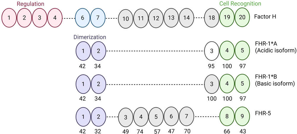
These original studies illustrate three complementary research patterns: functional regulation and renal-surface deposition in C3 glomerulopathy, a urinary complement regulator associated with IgA nephropathy progression, and paired plasma–urine complement profiling in FSGS.

Factor H and factor H-related protein domain architecture analyzed in C3 glomerulopathy research
C3G / FH–FHR balance / renal surface regulation

FHR-1 and heparan-sulfate architecture shape complement dysregulation in C3G

Slagle and colleagues combined patient FH/FHR measurements, genotype analysis, renal-cell C3b deposition, and heparan-sulfate binding studies. The work demonstrates how circulating regulator balance and local surface chemistry can be evaluated in one disease-mechanism program.

View research via DOI
CC BY 4.0
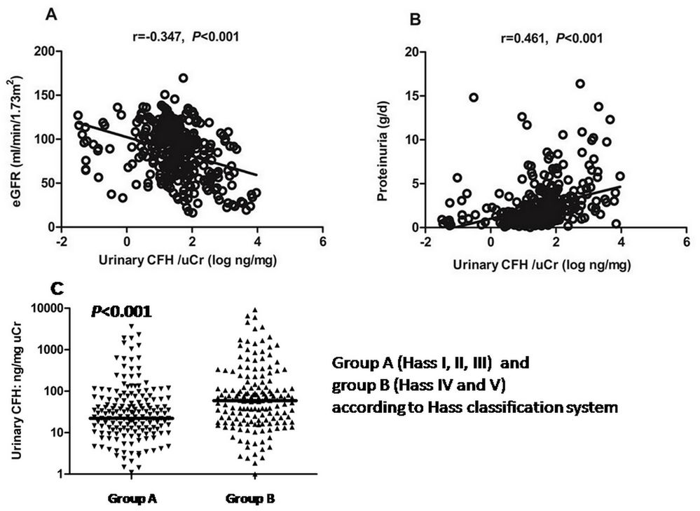
Urinary complement factor H relationships with renal function, proteinuria, and histologic changes in IgA nephropathy
IgA nephropathy / urinary CFH / progression

Urinary factor H connects complement regulation with IgAN severity and outcome

Liu and colleagues measured urinary CFH in a longitudinal IgA nephropathy cohort and related it to eGFR, proteinuria, histologic severity, and renal outcome. The design illustrates the value of matrix-aware complement biomarkers combined with established renal measures.

View research via DOI
CC BY
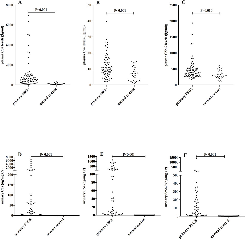
Plasma and urinary C3a, C5a, and soluble C5b-9 levels in primary focal segmental glomerulosclerosis
FSGS / plasma and urine / activation profile

Paired plasma and urinary complement profiles reveal pathway activity in FSGS

Huang and colleagues quantified C3a, C5a, sC5b-9, C4d, C1q, MBL, and Bb in biopsy-proven primary FSGS. The study shows how multi-analyte profiling can connect complement activation with proteinuria, renal dysfunction, fibrosis, treatment response, and outcome.

View research via DOI
CC BY
Questions and Answers

Frequently Asked Questions

What nephropathy and complement therapeutic research services does Creative Biolabs provide?

We provide complement pathway and activation-product profiling, C3 and C5b-9 deposition assays, functional genetic and autoantibody studies, renal cell and tissue models, serum/plasma/urine biomarker analysis, and complement-targeted candidate evaluation for glomerular, vascular, tubular, and transplant-related research contexts.

Which nephropathy research programs can be supported?

Programs can be designed for C3 glomerulopathy, complement-mediated HUS and thrombotic microangiopathy, IgA nephropathy, lupus nephritis, membranous nephropathy, other immune-complex glomerulonephritides, FSGS-related complement questions, tubular injury, and transplant-associated renal injury.

Can serum, plasma, urine, biopsy, and renal cell data be integrated?

Yes, subject to sample suitability and project requirements. A study can pair soluble pathway activity with urinary biomarkers, tissue deposition, renal cell phenotype, genotype, and candidate response. Collection and processing variables are reviewed because ex vivo complement activation and urine concentration can materially affect interpretation.

Can complement gene variants or autoantibodies be functionally evaluated?

Yes. Genetic findings and suspected autoantibodies can be linked to protein expression, binding, factor H or factor I regulation, convertase stability, C3b degradation, pathway output, renal-surface deposition, endothelial or renal-cell injury, and rescue by a functional regulator or inhibitor.

Which complement biomarkers are useful in nephropathy studies?

Options include C3a, C5a, Ba/Bb, iC3b/C3d, sC5b-9, C3, C4, C1q, factor B, factor D, properdin, factor H, factor I, FHR proteins, C4d, pathway functional activity, and C3-fragment or membrane attack complex deposition. The panel is selected according to disease mechanism and matrix.

Can complement inhibitors be evaluated in kidney-relevant models?

Yes. Candidates can be tested for target engagement, pathway selectivity, activation-product reduction, renal-surface deposition, endothelial or podocyte protection, permeability, inflammatory and thrombotic outputs, and toxicity in cell, co-culture, organoid, explant, or project-defined preclinical models.

What information is needed to design a customized nephropathy study?

Please provide the renal disease context, suspected complement pathway or target, available sample and species, kidney compartment, candidate format and amount, concentration range, benchmark, biomarker and phenotype endpoints, timeline, and the decision the data must support. Unknown items can be addressed in a feasibility phase.


Scientific Literature

References

  1. Slagle, Amanda K., et al. “Factor H-Related 1 and Heparan Sulfate Architecture Contribute to Complement Dysregulation in C3 Glomerulopathy.” Frontiers in Immunology, vol. 16, 2025, article 1589674. https://doi.org/10.3389/fimmu.2025.1589674
  2. Liu, Maojing, et al. “Implication of Urinary Complement Factor H in the Progression of Immunoglobulin A Nephropathy.” PLOS ONE, vol. 10, no. 6, 2015, e0126812. https://doi.org/10.1371/journal.pone.0126812
  3. Huang, Jing, et al. “Complement Activation Profile of Patients with Primary Focal Segmental Glomerulosclerosis.” PLOS ONE, vol. 15, no. 6, 2020, e0234934. https://doi.org/10.1371/journal.pone.0234934

Download

Online Inquiry