
Mechanism Mapping
Locate the pathway, compartment, surface, and disease phase where control is lost.


Creative Biolabs provides complement-focused AMD research services for programs investigating genetic susceptibility, alternative-pathway dysregulation, drusen-associated complement deposition, RPE and photoreceptor injury, geographic atrophy, choroidal neovascularization, translational biomarkers, and complement-targeted therapeutic candidates. Support spans study design, variant-to-function analysis, retinal and vascular models, pathway assays, efficacy testing, and integrated data interpretation.
Discuss Your AMD Research ProgramCreative Biolabs supports AMD programs in which complement is a causal hypothesis, a patient-stratification variable, a pharmacodynamic pathway, or a therapeutic target. Projects can begin with a risk variant in CFH, CFI, C3, CFB, or the CFHR locus; a retinal model showing complement-dependent stress; an ocular or circulating biomarker set; or an antibody, protein, peptide, aptamer, small molecule, or gene-based candidate.
We help translate that starting point into a staged research plan that aligns the complement source, retinal compartment, disease phase, trigger, assay matrix, comparator, candidate exposure, and success criteria. Studies are suitable for early target discovery, functional genetics, dry AMD and geographic atrophy research, neovascular AMD mechanism studies, biomarker development, lead selection, and preclinical proof of concept.
Programs may combine complement genetic testing, complement function and activity assays, retinal cell models, deposition analysis, angiogenesis readouts, and candidate-response testing.
Locate the pathway, compartment, surface, and disease phase where control is lost.
Test whether AMD-associated variants alter expression, binding, cofactor activity, or C3 regulation.
Connect complement activity to RPE, photoreceptor, immune, endothelial, and tissue outcomes.
Rank candidates by target engagement, pathway correction, retinal protection, and selectivity.
We can begin with pathway, activation-product, and deposition profiling before committing to a target-specific efficacy package.
AMD is heterogeneous: a complement risk allele does not define the complete phenotype, and pathway activation can be systemic, locally produced, surface restricted, or stress dependent. We therefore build experiments around a mechanistic chain that can be challenged at each step.
Complement risk or protective variants, aging, oxidative stress, lipid accumulation, smoking-related stress, extracellular matrix changes, or project-defined inflammatory triggers.
Altered factor H or factor I activity, impaired surface recognition, increased alternative-pathway amplification, insufficient RPE protection, or changed complement production.
C3 fragments, terminal-complex deposition, drusen-associated proteins, RPE dysfunction, inflammatory signaling, immune-cell recruitment, and photoreceptor vulnerability.
Geographic atrophy, outer-retinal degeneration, choroidal endothelial activation, vascular leakage, neovascular lesion growth, or treatment-modifiable biomarker patterns.
Does the proposed variant change factor H or factor I function? Is systemic complement activity reflected in the retinal phenotype? Which complement product accumulates on RPE or matrix surfaces? Does a candidate preserve RPE viability without completely suppressing host-defense activity?
Wild-type and risk-variant proteins, complement-depleted and reconstituted serum, heat-inactivated serum, pathway-selective blockade, oxidative or lipid stress controls, anti-VEGF or complement benchmarks, and rescue with a functional regulator can be used to establish causality.
Modules can be used independently or assembled into a staged package. Complement sources, serum concentration, stress exposure, time points, candidate dosing, and comparators are aligned so molecular and phenotypic results remain interpretable.
Determine whether an AMD-associated variant changes regulator abundance or quality.
Quantify the interactions that control alternative-pathway amplification on retinal surfaces.
Measure the net effect of disease context or treatment across the cascade.
Resolve whether complement accumulates on RPE, matrix, endothelial, or model surfaces.
Connect pathway modulation to outputs relevant to atrophy or neovascular disease.
Link genotype, pathway markers, retinal phenotype, and candidate response.
Matched cell-health, heat-inactivated serum, depleted-serum, and reconstitution controls can be built into the same study.
Purified systems are useful for variant mechanism, whereas retinal protection and candidate efficacy require the appropriate cell surface, extracellular matrix, stressor, complement source, and disease endpoint. We advance models only after the preceding assay tier is qualified.
Wild-type and variant proteins, depleted and reconstituted sera, convertase components, modified surfaces, and defined matrices for direct molecular and pathway studies.
ARPE-19, primary or donor-derived RPE, iPSC-derived RPE, photoreceptor-related cells, oxidative or lipid stress, barrier models, and complement-dependent injury.
Endothelial migration, tube formation, permeability, RPE–endothelial interfaces, macrophage or microglial components, and cytokine or VEGF-associated readouts.
Retinal organoids, tissue explants, drusen- or matrix-relevant systems, geographic atrophy concepts, laser-induced CNV, and model-specific pharmacodynamic endpoints.
Complement competence, species compatibility, serum concentration, RPE maturity and polarity, extracellular matrix, oxidative or lipid stress, donor genotype, collection and storage, freeze–thaw history, ex vivo complement activation, candidate exposure, and the dynamic range of the selected retinal endpoint.
A candidate is advanced only when its effect can be attributed to a defined complement mechanism and connected to retinal protection or vascular control in the selected AMD context.
Factor H or factor I replacement, engineered regulators, surface-targeted control, or functional rescue of a risk variant.
C3, factor B, factor D, properdin, convertase, amplification-loop, or deposition-focused intervention.
C5, C5a/C5aR, membrane attack complex, C3a/C3aR, inflammatory signaling, or tissue-injury modulation.
Complement modulation with anti-VEGF, antioxidant, anti-inflammatory, neuroprotective, or project-defined standard-of-care comparators.
Antibodies, recombinant proteins, peptides, aptamers, small molecules, gene-silencing tools, editing concepts, and local-delivery strategies can be compared using a milestone-based design.
Study controls may include vehicle, inactive analog, isotype, heat-inactivated serum, depleted and reconstituted serum, pathway-selective benchmarks, cell-health controls, and anti-angiogenic comparators.
Complement antibodies, proteins, inhibitors, and serum or plasma products can support controls, reconstitution, and candidate screening.
These original studies illustrate how complement variants can be tested beyond in silico prediction and how circulating pathway markers can be integrated with AMD-associated genetic information.
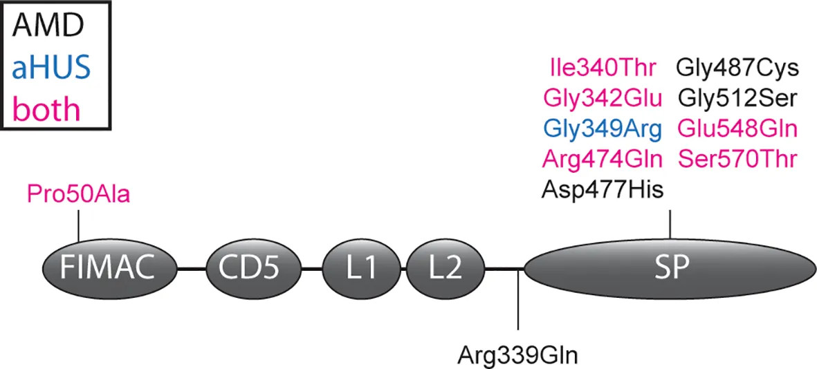
De Jong and colleagues expressed 11 rare CFI missense variants and quantified C3b degradation and iC3b generation. The study demonstrates how variants with apparently normal secretion can still produce a measurable loss of regulatory function.
View research via DOI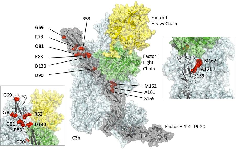
Wong and colleagues compared six CFH variants using C3b binding, surface plasmon resonance, cofactor assays, decay-acceleration studies, and hemolytic assays. Results separated a severe loss-of-function variant from subtler effects relevant to chronic complement-driven disease.
View research via DOI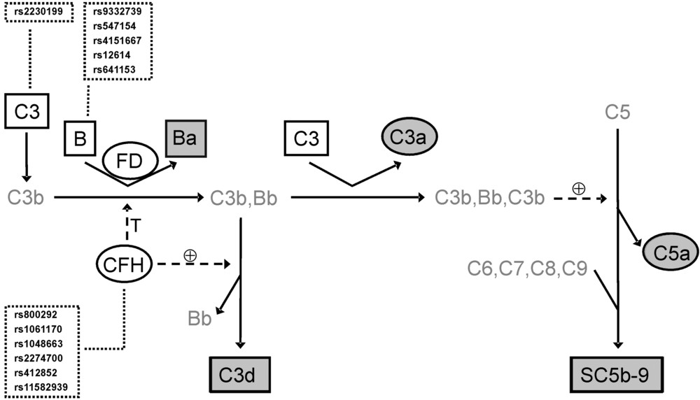
Scholl and colleagues measured activation products and complement proteins in AMD patients and controls while analyzing CFH, BF-C2, and C3 genetic markers. Ba, C3d, factor D, and other activation products supported an integrated biomarker–genotype strategy.
View research via DOIWe provide complement genetic and functional studies, factor H and factor I characterization, pathway and activation-product testing, C3 and C5b-9 deposition assays, RPE and retinal cell studies, endothelial and angiogenesis assays, biomarker profiling, AMD model development, and complement-targeted candidate evaluation.
Yes. Geographic atrophy-related programs can focus on chronic RPE or photoreceptor stress, complement deposition, inflammatory signaling, barrier function, and cell protection. Neovascular programs can add choroidal endothelial migration, tube formation, permeability, VEGF-associated signaling, macrophage responses, and lesion-related endpoints.
Yes. Wild-type and variant proteins can be compared for expression, secretion, stability, C3b or surface binding, factor I cofactor activity, C3b degradation, decay acceleration, pathway output, deposition, and rescue. The exact panel depends on the affected domain and proposed mechanism.
Options include Ba/Bb, C3a, C5a, C3d, sC5b-9, C3, C4, factor B, factor D, factor H, factor I, pathway functional activity, C3-fragment deposition, and membrane attack complex deposition. The panel is selected according to sample type, disease question, and candidate mechanism.
Yes, subject to sample suitability, availability, consent, and project requirements. Feasibility planning considers collection and processing, anticoagulant, storage, freeze–thaw history, ex vivo complement activation, matrix interference, donor genotype, cell maturity, sample volume, and the controls required for interpretation.
Yes. Complement candidates can be evaluated alone or with anti-angiogenic, antioxidant, anti-inflammatory, neuroprotective, or project-defined comparators. Studies can distinguish target engagement, complement pharmacodynamics, retinal protection, vascular response, additivity, and nonspecific toxicity.
Please provide the AMD stage or model, proposed complement target or genetic finding, available sample and species, retinal compartment, candidate format and amount, preferred concentration range, benchmark, required biomarkers and phenotype endpoints, timeline, and the decision the study must support. Unknown items can be resolved in a feasibility phase.

Complement Activity/Function Assay Products
Learn More
Complement Testing Services
Learn More
Complement Therapeutics Featured Products
Learn More
Complement Therapeutics Services Brochure
Learn More
Aptamer Development PLATFORM
Learn More
ComPLETTM Hemolysis Assay Solutions
Learn More