
Variant Prioritization
Choose variants and comparators using domain, population, phenotype, inheritance, and pathway context.


Creative Biolabs provides research services that connect complement-gene variation with measurable pathway dysfunction and therapeutic response. Programs support rare and complex genetic disease studies that need variant-to-function evidence, complement pathway and biomarker profiling, patient- or engineered-cell models, mechanism-of-action analysis, and preclinical evaluation of complement-targeted candidates.
Discuss Your Genetic Complement StudyCreative Biolabs helps research teams determine whether a reported or newly identified variant changes complement protein abundance, secretion, binding, proteolytic activity, convertase control, surface protection, or terminal-pathway injury. The work can start from a curated gene list, a variant of uncertain significance, a patient-derived matrix, an engineered construct, or a complement-targeted therapeutic hypothesis.
Services are suited to target discovery, genotype–phenotype investigation, functional variant classification, biomarker strategy, translational model development, mechanism-of-action studies, lead comparison, and preclinical proof of concept. Rather than treating genetics and complement testing as separate workstreams, we align molecular evidence, pathway activity, disease phenotype, and candidate response around the decision your program must make.
Study plans can combine complement genetic testing, recombinant-variant production, complement function and activity testing, activation-fragment analysis, cell-based assays, and disease-relevant models.
Choose variants and comparators using domain, population, phenotype, inheritance, and pathway context.
Measure expression, interaction, regulation, cleavage, deposition, and pathway consequences.
Reproduce the defect in serum, cell, organoid, tissue, or in vivo systems with matched controls.
Rank candidates by target engagement, pathway correction, phenotype rescue, and selectivity.
We can design a staged study that starts with protein and pathway confirmation before moving into complex disease models.
A pathogenic phenotype may arise from loss of a regulator, gain of an activator, impaired cell-surface protection, defective classical-pathway clearance, or a risk haplotype that becomes important only in a particular tissue or trigger context. Study design therefore follows the disease mechanism rather than a fixed test menu.
Evaluate CFH, CFI, CD46, C3, CFB, DGKE, and related findings in studies of alternative-pathway overactivation, endothelial injury, penetrance, trigger dependence, and response to terminal or proximal complement blockade.
Explore hemolytic uremic syndrome researchConnect variants or CFHR rearrangements with C3 consumption, convertase stability, factor H or factor I function, C3 fragment deposition, terminal activation, and renal-cell or matrix injury.
View C3b functional testingInvestigate CFH, CFI, C3, CFB, and CFHR risk or protective variants using binding, cofactor, cleavage, deposition, retinal-cell stress, and candidate-response assays relevant to complement-associated retinal pathology.
Explore factor H functional testingSupport SERPING1-centered research with antigenic and functional C1 inhibitor measurements, classical-pathway context, variant expression studies, and candidate rescue or replacement experiments.
Explore C1 inhibitor functional testingModel how PIGA or related defects alter CD55/CD59 display, C3-fragment deposition, membrane attack complex formation, complement-dependent cytotoxicity, hemolysis, and rescue by pathway-selective candidates.
View C5b-9 deposition assaysStudy classical-pathway deficiencies, infection susceptibility, immune-complex clearance, autoimmune predisposition, risk haplotypes, modifier alleles, or project-defined disorders where genetic background shapes complement response.
Explore autoimmune disease researchVariant studies are strongest when each experimental layer resolves a specific uncertainty. We select the shortest evidence path that can distinguish a true functional defect from altered expression, matrix artifacts, background genetics, or an unrelated disease process.
Define inheritance, domain location, conservation, allele frequency, phenotype concordance, and the reference sequence or isoform.
Assess expression, secretion, stability, processing, multimerization, and interaction with complement partners or surfaces.
Measure cofactor, decay acceleration, convertase, proteolysis, activation fragments, deposition, hemolysis, or receptor signaling.
Introduce the variant into relevant cells or proteins and apply triggers, serum matrices, patient samples, or tissue-specific stress.
Evaluate rescue by wild-type protein, reconstitution, pathway blockade, receptor modulation, gene-based tools, or lead candidates.
Protein generation, binding analysis, and functional assays can be coordinated under one experimental plan.
Modules can be used independently for focused confirmation or linked into a tiered program. Concentrations, matrices, incubation times, complement sources, and controls are harmonized so results can be interpreted across assay levels.
Determine whether the variant changes the amount or biochemical state of the complement component.
Quantify interactions that control amplification, convertase activity, ligand recognition, or surface protection.
Resolve loss or gain of regulatory activity beyond predicted structural impact.
Measure the net effect of a variant or candidate across classical, lectin, alternative, and terminal pathways.
Test whether genetically altered control changes opsonization or membrane attack on disease-relevant surfaces.
Align molecular findings with patient matrices, clinical phenotypes, triggers, and treatment-response hypotheses.
The same variant may appear neutral in a purified system yet become important on a vulnerable cell surface, under inflammatory stress, or in a serum background carrying additional risk alleles. Model selection therefore follows the proposed mechanism and the level of evidence required.
Wild-type and variant proteins, defined complement components, depleted sera, convertase assemblies, and biochemical surfaces for controlled mechanism studies.
Gene-edited lines, transfected cells, endothelial cells, erythroid cells, renal or retinal cells, iPSC-derived systems, and primary immune cells.
Cell–cell interfaces, tissue-specific stress, extracellular matrix, organoid platforms, explants, and deposition-compatible surfaces.
Model selection around complement genotype, species biology, disease trigger, tissue outcome, candidate exposure, pharmacodynamics, and rescue endpoints.
A feasibility phase can identify matrix effects and establish the most informative model sequence before scarce samples are committed.
Candidate testing is designed to separate biochemical target engagement from pathway correction and true phenotype rescue. The result is a traceable decision path rather than a single inhibition value.
Confirm binding, competition, component inhibition, receptor occupancy, or replacement of the missing activity.
Measure activation fragments, convertase control, deposition, hemolysis, or terminal-complex suppression in the variant context.
Demonstrate restoration of cell protection, endothelial behavior, tissue response, inflammatory balance, or disease-model outcome.
Compare potency with cell health, residual host-defense activity, matrix dependence, donor variability, and benchmark performance.
Programs can compare complement-specific antibodies, recombinant regulators, replacement proteins, peptides, aptamers, small molecules, receptor antagonists, gene-silencing tools, editing concepts, or combination strategies.
Candidate discovery and assay development can also be combined through our complement component inhibitor development capabilities.
Browse complement antibodies, proteins, inhibitors, and serum or plasma products for controls, reconstitution, and screening.
Each stage has a defined question, quality check, and progression criterion. Programs can stop after focused functional confirmation or continue through model development and therapeutic evaluation.
Define the gene, variant, inheritance or clone context, phenotype, proposed pathway effect, available material, comparator, candidate, and required decision.
Confirm reagent quality, matrix compatibility, dynamic range, complement activity, expression level, controls, and sample requirements.
Run matched wild-type and variant studies across expression, binding, regulation, pathway activity, deposition, or cell-protection endpoints.
Transfer the validated defect into the selected cell, tissue, or preclinical model; establish dose, timing, pharmacodynamics, rescue, and benchmark comparison.
Deliver methods, QC, raw and processed data, statistical outputs, figures, variant-specific interpretation, candidate ranking, and next-step recommendations.
The selected research demonstrates why biochemical and pathway assays are needed after variant discovery, and how functional data can distinguish severe loss of regulation from subtle or context-dependent effects.
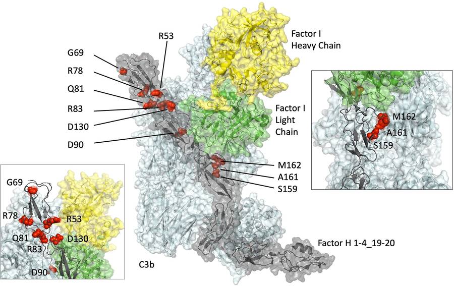
Wong and colleagues compared six CFH variants using protein production, C3b binding, surface plasmon resonance, cofactor assays, and hemolytic assays. The study illustrates how experimental evidence can separate a profound defect from subtle or undetectable functional changes.
View research via DOI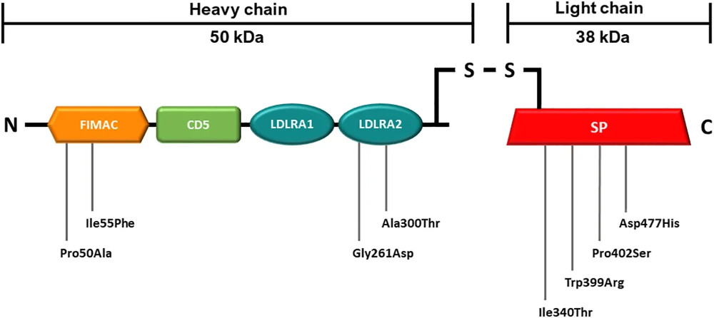
Gerogianni and colleagues assessed eight recombinant factor I variants with SDS-PAGE, ELISA, and a Luminex bead-based surface assay using factor H or soluble CR1 as cofactors. The work shows why assay context and multiple readouts matter when variant effects are modest.
View research via DOI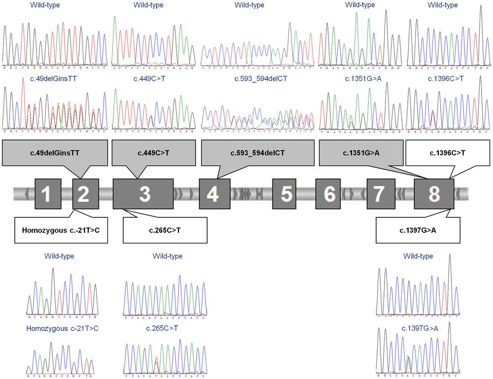
Rijavec and colleagues combined clinical and complement measurements with sequencing and copy-number analysis in families with hereditary angioedema. The study highlights genetic heterogeneity and the value of pairing a molecular finding with C1 inhibitor level and function.
View research via DOIWe provide variant-to-function study design, recombinant wild-type and variant protein work, expression and binding analysis, complement component and pathway assays, activation-product and deposition testing, cell-based phenotype studies, disease-model development, biomarker profiling, and complement-targeted candidate evaluation.
Yes. The study can compare the variant with wild-type, benign, and known loss-of-function controls across expression, secretion, interaction, regulatory activity, pathway output, deposition, and cell-protection assays. The final panel depends on the protein domain, proposed mechanism, disease context, and available evidence.
Programs may address CFH, CFI, C3, CFB, CD46, SERPING1, CFHR genes, PIGA-related surface protection, classical-pathway deficiencies, or other project-defined genes. Mechanisms can include quantitative deficiency, impaired cofactor or decay activity, convertase stabilization, altered receptor signaling, defective membrane protection, or abnormal terminal-pathway activation.
Yes. Complement genetic testing can be linked with protein-level analysis, pathway activity, activation products, deposition, hemolysis, receptor or ligand interaction, and disease-relevant cell assays. This integrated approach helps determine whether a genetic finding has a measurable biological consequence.
Yes, subject to sample suitability, consent, and project requirements. Feasibility planning considers anticoagulant, processing time, storage, freeze–thaw history, ex vivo complement activation, matrix interference, sample volume, and the controls required to distinguish genotype-associated effects from pre-analytical variation.
Yes. Candidates can be assessed for target engagement, pathway correction, deposition or hemolysis inhibition, phenotype rescue, selectivity, and dose response in matched wild-type and variant systems. Rescue with wild-type protein or pathway-selective controls can strengthen causal interpretation.
Please share the gene and variant notation, reference transcript, disease phenotype, inheritance or clone context, prior genetic and complement data, available samples or constructs, preferred model, candidate information, benchmark, timeline, and the decision the study must support. If the mechanism is uncertain, we can begin with a focused feasibility phase.

Complement Activity/Function Assay Products
Learn More
Complement Testing Services
Learn More
Complement Therapeutics Featured Products
Learn More
Complement Therapeutics Services Brochure
Learn More
Aptamer Development PLATFORM
Learn More
ComPLETTM Hemolysis Assay Solutions
Learn More