Research Highlights

Topics

All Topics

By Complement Pathways

Alternative Pathway

Classical Pathway

Lectin Pathway

Membrane Attack Complex

Terminal Pathway

By Diseases

Cardiovascular Diseases

Infectious Diseases

Inflammatory Diseases

Renal Diseases

Neurological Diseases

RheumaticDiseases

Other Diseases

By Applications

Therapeutics

Diagnostics

Vaccines

Others

photo_0_pro.png

Exposure to particulate matter (PM) is becoming a major global health issue. The amount and time of exposure to PM are known to be closely associated with cardiovascular diseases. However, the mechanism through which PM affects the vascular system is still not clear. Endothelial cells line the interior surface of blood vessels and actively interact with plasma proteins, including the complement system. Unregulated complement activation caused by invaders, such as pollutants, may promote endothelial inflammation. In the present study, we sought to investigate whether urban PM (UPM) acts on the endothelial environment via the complement system. UPM-treated human endothelial cells with normal human serum showed the deposition of membrane attack complexes (MACs) on the cell surface via the alternative pathway of the complement system. Despite the formation of MACs, cell death was not observed, and cell proliferation was increased in UPM-mediated complement activation. Furthermore, complement activation on endothelial cells stimulated the production of inflammation-related proteins. Our results revealed that UPM could activate the complement system in human endothelial cells and that complement activation regulated inflammatory reaction in microenvironment. These findings provide clues with regard to the role of the complement system in pathophysiologic events of vascular disease elicited by air pollution.
Keywords: complement system; endothelial cell; inflammation; particulate matter.

photo_1_pro.png

Objective: The concentrations of complement proteins (adipsin, C3a, and C5a) and soluble endoglin (sENG) in the plasma were measured in this study, and their value as early-pregnancy predictors and potential diagnostic marker of preeclampsia was assessed, respectively.
Experimental design: Plasma samples were obtained from healthy and preeclampsia pregnant women before delivery for a cross-sectional study. Plasma samples were collected from healthy and preeclampsia pregnant women throughout pregnancy and postpartum for a follow-up study. Enzyme-linked immunosorbent assays were used to detect plasma levels of several complement proteins (adipsin, C3a, and C5a) and sENG.
Results: The plasma levels of adipsin, C5a, and sENG were significantly increased before delivery in pregnant women with preeclampsia. During pregnancy, the plasma adipsin, C5a, and sENG levels were increased from the third trimester in healthy pregnant women; plasma adipsin levels remained stable after delivery, while C3a levels increased in the second trimester and remained stable afterward. Furthermore, levels of adipsin, C5a, and sENG were higher in preeclampsia patients at different stages of pregnancy; the C3a level presents a similar change and no difference was found in the third trimester. In the first trimester, receiver-operating curve (ROC) curve analysis showed that adipsin (AUC, 0.83 ± 0.06, P=0.001) and sENG (AUC, 0.74 ± 0.09, P=0.021) presented high value as predictors of early pregnancy.
Conclusions: Adipsin is likely a novel plasma biomarker to monitor the increased risk of preeclampsia in early pregnancy. Moreover, the increased plasma levels of adipsin, C5a, and sENG before delivery may be associated with preeclampsia.
Keywords: adipsin; biomarker; complement system; preeclampsia; sENG.

photo_2_pro.png

Familial hypercholesterolemia (FH) is increasingly associated with inflammation, a phenotype that persists despite treatment with lipid lowering therapies. The alternative C3 complement system (C3), as a key inflammatory mediator, seems to be involved in the atherosclerotic process; however, the relationship between C3 and lipids during plaque progression remains unknown. The aim of the study was to investigate by a systems biology approach the role of C3 in relation to lipoprotein levels during atherosclerosis (AT) progression and to gain a better understanding on the effects of C3 products on the phenotype and function of human lipid-loaded vascular smooth muscle cells (VSMCs). By mass spectrometry and differential proteomics, we found the extracellular matrix (ECM) of human aortas to be enriched in active components of the C3 complement system, with a significantly different proteomic signature in AT segments. Thus, C3 products were more abundant in AT-ECM than in macroscopically normal segments. Furthermore, circulating C3 levels were significantly elevated in FH patients with subclinical coronary AT, evidenced by computed tomographic angiography. However, no correlation was identified between circulating C3 levels and the increase in plaque burden, indicating a local regulation of the C3 in AT arteries. In cell culture studies of human VSMCs, we evidenced the expression of C3, C3aR (anaphylatoxin receptor) and the integrin αMβ2 receptor for C3b/iC3b (RT-PCR and Western blot). C3mRNA was up-regulated in lipid-loaded human VSMCs, and C3 protein significantly increased in cell culture supernatants, indicating that the C3 products in the AT-ECM have a local vessel-wall niche. Interestingly, C3a and iC3b (C3 active fragments) have functional effects on VSMCs, significantly reversing the inhibition of VSMC migration induced by aggregated LDL and stimulating cell spreading, organization of F-actin stress fibers and attachment during the adhesion of lipid-loaded human VSMCs. This study, by using a systems biology approach, identified molecular processes involving the C3 complement system in vascular remodeling and in the progression of advanced human atherosclerotic lesions.
Keywords: atherosclerosis; cardiovascular disease; complement system; mass spectrometry; proteomics.

photo_3_pro.png

Antimicrobial peptides (AMPs) are not only cytotoxic towards host pathogens or cancer cells but also are able to act as immunomodulators. It was shown that some human and non-human AMPs can interact with complement proteins and thereby modulate complement activity. Thus, AMPs could be considered as the base for complement-targeted therapeutics development. Arenicins from the sea polychaete Arenicola marina, the classical example of peptides with a β-hairpin structure stabilized by a disulfide bond, were shown earlier to be among the most prospective regulators. Here, we investigate the link between arenicins' structure and their antimicrobial, hemolytic and complement-modulating activities using the derivative Ar-1-(C/A) without a disulfide bond. Despite the absence of this bond, the peptide retains all important functional activities and also appears less hemolytic in comparison with the natural forms. These findings could help to investigate new complement drugs for regulation using arenicin derivatives.

photo_5_pro.png

Neuromyelitis optica spectrum disorder (NMOSD) is an autoimmune inflammatory disease of the central nervous system (CNS), characterized by pathogenic, complement-activating autoantibodies against the main water channel in the CNS, aquaporin 4 (AQP4). NMOSD is frequently associated with additional autoantibodies and antibody-mediated diseases. Because the alternative pathway amplifies complement activation, our aim was to evaluate the presence of autoantibodies against the alternative pathway C3 convertase, its components C3b and factor B, and the complement regulator factor H (FH) in NMOSD. Four out of 45 AQP4-seropositive NMOSD patients (~9%) had FH autoantibodies in serum and none had antibodies to C3b, factor B and C3bBb. The FH autoantibody titers were low in three and high in one of the patients, and the avidity indexes were low. FH-IgG complexes were detected in the purified IgG fractions by Western blot. The autoantibodies bound to FH domains 19-20, and also recognized the homologous FH-related protein 1 (FHR-1), similar to FH autoantibodies associated with atypical hemolytic uremic syndrome (aHUS). However, in contrast to the majority of autoantibody-positive aHUS patients, these four NMOSD patients did not lack FHR-1. Analysis of autoantibody binding to FH19-20 mutants and linear synthetic peptides of the C-terminal FH and FHR-1 domains, as well as reduced FH, revealed differences in the exact binding sites of the autoantibodies. Importantly, all four autoantibodies inhibited C3b binding to FH. In conclusion, our results demonstrate that FH autoantibodies are not uncommon in NMOSD and suggest that generation of antibodies against complement regulating factors among other autoantibodies may contribute to the complement-mediated damage in NMOSD.
Keywords: aquaporin (AQP) 4; autoantibody; autoimmunity; central nervous system; complement; factor H; inflammation; neuromyelitis optica spectrum disorder.

photo_6_pro.png

C4b Binding Protein (C4BP) is a major fluid phase inhibitor of the classical and lectin pathways of the complement system. Complement inhibition is achieved by binding to and restricting the role of activated complement component C4b. C4BP functions as a co-factor for factor I in proteolytic inactivation of both soluble and cell surface-bound C4b, thus restricting the formation of the C3-convertase, C4b2a. C4BP also accelerates the natural decay/dissociation of the C3 convertase. This makes C4BP a prime target for exploitation by pathogens to escape complement attack, as seen in Streptococcus pyogenes or Flavivirus. Here, we examined whether C4BP can act on its own in a complement independent manner, against pathogens. C4BP bound H1N1 and H3N2 subtypes of Influenza A Virus (IAV) most likely via multiple sites in Complement Control Protein (CCP) 1-2, 4-5, and 7-8 domains of its α-chain. In addition, C4BP CCP1-2 bound H3N2 better than H1N1. C4BP bound three IAV envelope proteins: Haemagglutinin (~70 kDa), Neuraminidase (~55 kDa), and Matrix protein 1 (~25kDa). C4BP suppressed H1N1 subtype infection into the lung epithelial cell line, A549, while it promoted infection by H3N2 subtype. C4BP restricted viral entry for H1N1 but had the opposite effect on H3N2, as evident from experiments using pseudo-typed viral particles. C4BP downregulated mRNA levels of pro-inflammatory IFN-α, IL-12, and NFκB in the case of H1N1, while it promoted a pro-inflammatory immune response by upregulating IFN- α, TNF-α, RANTES, and IL-6 in the case of H3N2. We conclude that C4BP differentially modulates the efficacy of IAV entry, and hence, replication in a target cell in a strain-dependent manner, and acts as an entry inhibitor for H1N1. Thus, CCP containing complement proteins such as factor H and C4BP may have additional defense roles against IAV that do not rely on the regulation of complement activation.
Keywords: C4BP; complement; inflammation; influenza A virus; pseudo-typed lentiviral particles.

photo_8_pro.png

Introduction: Multiple sclerosis (MS) is a demyelinating disease of the central nervous system with an underlying immune-mediated and inflammatory pathogenesis. Innate immunity, in addition to the adaptive immune system, plays a relevant role in MS pathogenesis. It represents the immediate non-specific defense against infections through the intrinsic effector mechanism “immunothrombosis” linking inflammation and coagulation. Moreover, decreased cerebral blood volume (CBV), cerebral blood flow (CBF), and prolonged mean transit time (MTT) have been widely 
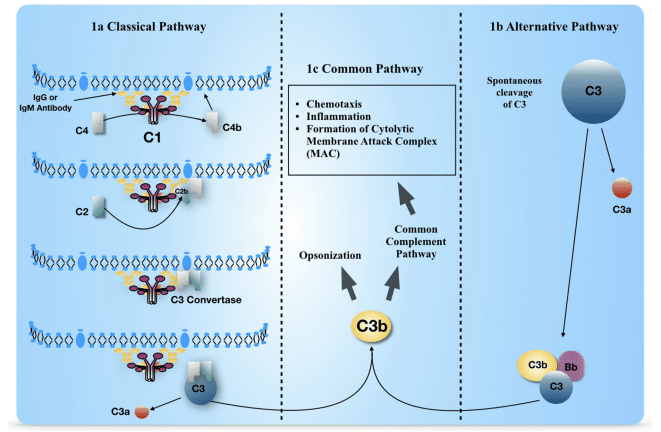
demonstrated by MRI in MS patients. We hypothesized that coagulation/complement and platelet activation during MS relapse, likely during viral infections, could be related to CBF decrease. Our specific aims are to evaluate whether there are differences in serum/plasma levels of coagulation/complement factors between relapsing-remitting (RR) MS patients (RRMS) in relapse and those in remission and healthy controls as well as to assess whether brain hemodynamic changes detected by MRI occur in relapse compared with remission. This will allow us to correlate coagulation status with perfusion and demographic/clinical features in MS patients.
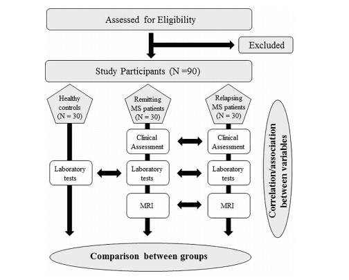
Materials and Methods: This is a multi-center, prospective, controlled study. RRMS patients (1° group: 30 patients in relapse; 2° group: 30 patients in remission) and age/sex-matched controls (3° group: 30 subjects) will be enrolled in the study. Patients and controls will be tested for 
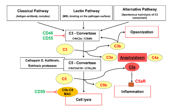
either coagulation/complement (C3, C4, C4a, C9, PT, aPTT, fibrinogen, factor II, VIII, and X, D-dimer, antithrombin, protein C, protein S, von-Willebrand factor), soluble markers of endothelial damage (thrombomodulin, Endothelial Protein C Receptor), antiphospholipid antibodies, lupus anticoagulant, complete blood count, viral serological assays, or microRNA microarray. Patients will undergo dynamic susceptibility contrast-enhanced MRI using a 3.0-T scanner to evaluate CBF, CBV, MTT, lesion number, and volume.
Statistical Analysis: ANOVA and unpaired t-tests will be used. The level of significance was set at p ≤ 0.05.
Discussion: Identifying a link between activation of coagulation/complement system and cerebral hypoperfusion could improve the identification of novel molecular and/or imaging biomarkers and targets, leading to the development of new effective therapeutic strategies in MS.
Clinical Trial Registration: Clinicaltrials.gov, identifier NCT04380220.
Keywords: multiple sclerosis, coagulation, complement, platelets, relapse, infection, cerebral hypoperfusion

photo_9_pro.png

Complement, a feature of the innate immune system that targets pathogens for phagocytic clearance and promotes inflammation, is tightly regulated to prevent damage to host tissue. This regulation is paramount in the central nervous system (CNS) since complement proteins degrade neuronal synapses during development, homeostasis, and neurodegeneration. We propose that dysregulated complement, particularly C1 or C3b, may errantly target synapses for immune-mediated clearance, therefore highlighting regulatory failure as a major potential mediator of neurological disease. First, we explore the mechanics of molecular neuroimmune relationships for the regulatory proteins: Complement Receptor 1, C1-Inhibitor, Factor H, and the CUB-sushi multiple domain family. We propose that biophysical and chemical principles offer clues for understanding mechanisms of dysregulation. Second, we describe anticipated effects to CNS disease processes (particularly Alzheimer's Disease) and nest our ideas within existing basic science, clinical, and epidemiological findings. Finally, we illustrate how the concepts presented within this manuscript provoke new ways of approaching age-old neurodegenerative processes. Every component of this model is testable by straightforward experimentation and highlights the untapped potential of complement dysregulation as a driver of CNS disease. This includes a putative role for complement-based neurotherapeutic agents and companion biomarkers.
Keywords: Alzheimer's disease; C1 inhibitor; CR1; complement; factor H; neuroimmune; schizophrenia.

photo_11_pro.png

Background: The SARS-CoV-2 infection triggers excessive immune response resulting in increased levels of pro-inflammatory cytokines, endothelial injury, and intravascular coagulopathy. The complement system (CS) activation participates to this hyperinflammatory response. However, it is still unclear which activation pathways (classical, alternative, or lectin pathway) pilots the effector mechanisms that contribute to critical illness. To better understand the immune correlates of disease severity, we performed an analysis of CS activation pathways and components in samples collected from COVID-19 patients hospitalized in Grenoble Alpes University Hospital between 1 and 30 April 2020 and of their relationship with the clinical outcomes.
Methods: We conducted a retrospective, single-center study cohort in 74 hospitalized patients with RT-PCR-proven COVID-19. The functional activities of classical, alternative, and mannose-binding lectin (MBL) pathways and the antigenic levels of the individual components C1q, C4, C3, C5, Factor B, and MBL were measured in patients' samples during hospital admission. Hierarchical clustering with the Ward method was performed in order to identify clusters of patients with similar characteristics of complement markers. Age was included in the model. Then, the clusters were compared with the patient clinical features: rate of intensive care unit (ICU) admission, corticoid treatment, oxygen requirement, and mortality.
Results: Four clusters were identified according to complement parameters. Among them, two clusters revealed remarkable profiles: in one cluster (n = 15), patients exhibited activation of alternative and lectin pathways and low antigenic levels of MBL, C4, C3, Factor B, and C5 compared to all the other clusters; this cluster had the higher proportion of patients who died (27%) and required oxygen support (80%) or ICU care (53%). In contrast, the second cluster (n = 19) presented inflammatory profile with high classical pathway activity and antigenic levels of complement components; a low proportion of patients required ICU care (26%) and no patient died in this group.
Conclusion: These findings argue in favor of prominent activation of the alternative and MBL complement pathways in severe COVID-19, but the spectrum of complement involvement seems to be heterogeneous requiring larger studies.
Keywords: COVID-19; MBL; alternative pathway; complement; lectin pathway.

photo_13_pro.png

In recent years, accumulating evidence has shown that the innate immune complement system is involved in several aspects of normal brain development and in neurodevelopmental disorders, including autism spectrum disorder (ASD). Although abnormal expression of complement components was observed in post-mortem brain samples from individuals with ASD, little is known about the expression patterns of complement molecules in distinct cell types in the developing autistic brain. In the present study, we characterized the mRNA and protein expression profiles of a wide range of complement system components, receptors and regulators in induced pluripotent stem cell (iPSC)-derived neural progenitor cells, neurons and astrocytes of individuals with ASD and neurotypical controls, which constitute in vitro cellular models that recapitulate certain features of both human brain development and ASD pathophysiology. We observed that all the analyzed cell lines constitutively express several key complement molecules. Interestingly, using different quantification strategies, we found that complement C4 mRNA and protein are expressed in significantly lower levels by astrocytes derived from ASD individuals compared to control astrocytes. As astrocytes participate in synapse elimination, and diminished C4 levels have been linked to defective synaptic pruning, our findings may contribute to an increased understanding of the atypically enhanced brain connectivity in ASD.
Keywords: autism spectrum disorder; complement C4; complement system; iPSC-derived astrocytes; synaptic pruning.

photo_15_pro.png

Clearance of viral infections, such as SARS-CoV-2 and influenza A virus (IAV), must be fine-tuned to eliminate the pathogen without causing immunopathology. As such, an aggressive initial innate immune response favors the host in contrast to a detrimental prolonged inflammation. The complement pathway bridges innate and adaptive immune system and contributes to the response by directly clearing pathogens or infected cells, as well as recruiting proinflammatory immune cells and regulating inflammation. However, the impact of modulating complement activation in viral infections is still unclear. In this work, we targeted the complement decay-accelerating factor (DAF/CD55), a surface protein that protects cells from non-specific complement attack, and analyzed its role in IAV infections. We found that DAF modulates IAV infection in vivo, via an interplay with the antigenic viral proteins hemagglutinin (HA) and neuraminidase (NA), in a strain specific manner. Our results reveal that, contrary to what could be expected, DAF potentiates complement activation, increasing the recruitment of neutrophils, monocytes and T cells. We also show that viral NA acts on the heavily sialylated DAF and propose that the NA-dependent DAF removal of sialic acids exacerbates complement activation, leading to lung immunopathology. Remarkably, this mechanism has no impact on viral loads, but rather on the host resilience to infection, and may have direct implications in zoonotic influenza transmissions.

photo_16_pro.png

A central part of the complement system, the anaphylatoxin C5a was investigated in this study to learn its effects on tenocytes in respect to understanding the potential expression of other crucial complement factors and pro-inflammatory mediators involved in tendinopathy. Human hamstring tendon-derived tenocytes were treated with recombinant C5a protein in concentrations of 25 ng/mL and 100 ng/mL for 0.5 h (early phase), 4 h (intermediate phase), and 24 h (late phase). Tenocytes survival was assessed after 24 h stimulation by live-dead assay. The gene expression of complement-related factors C5aR, the complement regulatory proteins (CRPs) CD46, CD55, CD59, and of the pro-inflammatory cytokines tumor necrosis factor (TNF)-α and interleukin (IL)-6 was monitored using qPCR. Tenocytes were immunolabeled for C5aR and CD55 proteins. TNFα production was monitored by ELISA. Tenocyte survival was not impaired through C5a stimulation. Interestingly, the gene expression of C5aR and that of the CRPs CD46 and CD59 was significantly reduced in the intermediate and late phase, and that of TNFα only in an early phase, compared to the control group. ELISA analysis indicated a concomitant not significant trend of impaired TNFα protein synthesis at 4 h. However, there was also an early significant induction of CD55 and CD59 mediated by 25 ng/mL anaphylatoxin C5a. Hence, exposure of tenocytes to C5a obviously evokes a time and concentration-dependent response in their expression of complement and pro-inflammatory factors. C5a, released in damaged tendons, might directly contribute to tenocyte activation and thereby be involved in tendon healing and tendinopathy.
Keywords: C5a; anaphylatoxin; complement regulation; complement system; tendinopathy; tenocytes.

photo_18_pro.png

Complement is an enzymatic humoral pattern-recognition defence system of the body. Non-specific deposition of blood biomolecules on nanomedicines triggers complement activation through the alternative pathway, but complement-triggering mechanisms of nanomaterials with dimensions comparable to or smaller than many globular blood proteins are unknown. Here we study this using a library of <6 nm poly(amido amine) dendrimers bearing different end-terminal functional groups. Dendrimers are not sensed by C1q and mannan-binding lectin, and hence do not trigger complement activation through these pattern-recognition molecules. While, pyrrolidone- and carboxylic acid-terminated dendrimers fully evade complement response, and independent of factor H modulation, binding of amine-terminated dendrimers to a subset of natural IgM glycoforms triggers complement activation through lectin pathway-IgM axis. These findings contribute to mechanistic understanding of complement surveillance of dendrimeric materials, and provide opportunities for dendrimer-driven engineering of complement-safe nanomedicines and medical devices.

photo_22_pro.png

Acute lung injury (ALI) is the injury of alveolar epithelial cells and capillary endothelial cells caused by various factors. Complement system and pyroptosis have been proved to be involved in ALI, and inhibition of C5a/C5a receptor (C5aR) could alleviate ALI. This study aimed to investigate whether C5a/C5aR inhibition could protect against LPS-induced ALI via mediating pyroptosis. Rats were assigned into four groups: Control, LPS, LPS+W-54011 1mg/kg, and LPS+W-54011 5mg/kg. Beas-2B cells pretreated with or without C5a and W-54011, alone and in combination, were challenged with LPS+ATP. Results unveiled that LPS caused lung tissue injury and inflammatory response, increased pyroptotic and apoptotic factors, along with elevated C5a concentration and C5aR expressions. However, W-54011 pretreatment alleviated lung damage and pulmonary edema, reduced inflammation and prevented cell pyroptosis. In vitro studies confirmed that LPS+ATP reduced cell viability, promoted cell death, generated inflammatory factors and promoted expressions of pyroptosis-related proteins, which could be prevented by W-54011 pretreatment while intensified by C5a pretreatment. The co-treatment of C5a and W-54011 could blunt the effects of C5a on LPS+ATP-induced cytotoxicity. In conclusion, inhibition of C5a/C5aR developed protective effects against LPS-induced ALI and the cytotoxicity of Beas-2B cells, and these effects may depend on blocking pyroptosis.
Keywords: C5a; acute lung injury; complement; inflammation; pyroptosis.

photo_23_pro.png

The complement system has developed different strategies to clear infections by several effector mechanisms, such as opsonization, which supports phagocytosis, attracting immune cells by C3 and C5 cleavage products, or direct killing of pathogens by the formation of the membrane attack complex (MAC). As the Zika virus (ZIKV) activates the classical complement pathway and thus has to avoid clearance by the complement system, we analyzed putative viral escape mechanisms, which limit virolysis. We identified binding of the recombinant viral envelope E protein to components of the terminal pathway complement (C5b6, C7, C8, and C9) by ELISA. Western blot analyses revealed that ZIKV E protein interfered with the polymerization of C9, induced on cellular surfaces, either by purified terminal complement proteins or by normal human serum (NHS) as a source of the complement. Further, the hemolytic activity of NHS was significantly reduced in the presence of the recombinant E protein or entire viral particles. This data indicates that ZIKV reduces MAC formation and complement-mediated lysis by binding terminal complement proteins to the viral E protein.
Keywords: complement, lysis, membrane attack complex, Zika virus, envelope protein, terminal complement pathway

photo_24_pro.png

Early and persistent activation of complement is considered to play a key role in the pathogenesis of COVID-19. Complement activation products orchestrate a proinflammatory environment that might be critical for the induction and maintenance of a severe inflammatory response to SARS-CoV-2 by recruiting cells of the cellular immune system to the sites of infection and shifting their state of activation towards an inflammatory phenotype. It precedes pathophysiological milestone events like the cytokine storm, progressive endothelial injury triggering microangiopathy, and further complement activation, and causes an acute respiratory distress syndrome (ARDS). To date, the application of antiviral drugs and corticosteroids have shown efficacy in the early stages of SARS-CoV-2 infection, but failed to ameliorate disease severity in patients who progressed to severe COVID-19 pathology. This report demonstrates that lectin pathway (LP) recognition molecules of the complement system, such as MBL, FCN-2 and CL-11, bind to SARS-CoV-2 S- and N-proteins, with subsequent activation of LP-mediated C3b and C4b deposition. In addition, our results confirm and underline that the N-protein of SARS-CoV-2 binds directly to the LP- effector enzyme MASP-2 and activates complement. Inhibition of the LP using an inhibitory monoclonal antibody against MASP-2 effectively blocks LP-mediated complement activation. FACS analyses using transfected HEK-293 cells expressing SARS-CoV-2 S protein confirm a robust LP-dependent C3b deposition on the cell surface which is inhibited by the MASP-2 inhibitory antibody. In light of our present results, and the encouraging performance of our clinical candidate MASP-2 inhibitor Narsoplimab in recently published clinical trials, we suggest that the targeting of MASP-2 provides an unsurpassed window of therapeutic efficacy for the treatment of severe COVID-19.
Keywords: COVID-19; SARS-CoV-2; complement system; innate immunity; lectin pathway.

photo_25_pro.png

Partial phagocytosis-called trogocytosis-of axons by microglia has been documented in ex vivo preparations but has not been directly observed in vivo. The mechanisms that modulate microglial trogocytosis of axons and its function in neural circuit development remain poorly understood. Here, we directly observe axon trogocytosis by microglia in vivo in the developing Xenopus laevis retinotectal circuit. We show that microglia regulate pruning of retinal ganglion cell axons and are important for proper behavioral response to dark and bright looming stimuli. Using bioinformatics, we identify amphibian regulator of complement activation 3, a homolog of human CD46, as a neuronally expressed synapse-associated complement inhibitory molecule that inhibits trogocytosis and axonal pruning. Using a membrane-bound complement C3 fusion protein, we demonstrate that enhancing complement activity enhances axonal pruning. Our results support the model that microglia remodel axons via trogocytosis and that neurons can control this process through expression of complement inhibitory proteins.
Keywords: axon; behavior; immunology; inflammation; microglia; morphometry; neuroscience; pruning; retinotectal; xenopus.

photo_26_pro.png

Pentraxin 3 (PTX3) is an essential component of the innate immune system and a recognized modulator of Complement cascade. The role of Complement system in the pathogenesis of prostate cancer has been largely underestimated. The aim of our study was to investigate the role of PTX3 as possible modulator of Complement activation in the development of this neoplasia. We performed a single center cohort study; from January 2017 through December 2018, serum and prostate tissue samples were obtained from 620 patients undergoing prostate biopsy. A group of patients with benign prostatic hyperplasia (BPH) underwent a second biopsy within 12-36 months demonstrating the presence of a prostate cancer (Group A, n = 40) or confirming the diagnosis of BPH (Group B, N = 40). We measured tissue PTX3 protein expression together with complement activation by confocal microscopy in the first and second biopsy in group A and B patients. We confirmed that that PTX3 tissue expression in the first biopsy was increased in group A compared to group B patients. C1q deposits were extensively present in group A patients co-localizing and significantly correlating with PTX3 deposits; on the contrary, C1q/PTX3 deposits were negative in group B. Moreover, we found a significantly increased expression of C3a and C5a receptors within resident cells in group A patient. Interestingly, C1q/PTX3 deposits were not associated with activation of the terminal Complement complex C5b-9; moreover, we found a significant increase of Complement inhibitor CD59 in cancer tissue. Our data indicate that PTX3 might play a significant pathogenic role in the development of this neoplasia through recruitment of the early components of Complement cascade with hampered activation of terminal Complement pathway associated with the upregulation of CD59. This alteration might lead to the PTX3-mediated promotion of cellular proliferation, angiogenesis and insensitivity to apoptosis possible leading to cancer cell invasion and migration.

photo_27_pro.png

Rituximab is a pioneering anti-CD20 monoclonal antibody that became the first-line drug used in immunotherapy of B-cell malignancies over the last twenty years. Rituximab activates the complement system in vitro, but there is an ongoing debate on the exact role of this effector mechanism in therapeutic effect. Results of both in vitro and in vivo studies are model-dependent and preclude clear clinical conclusions. Additional confounding factors like complement inhibition by tumor cells, loss of target antigen and complement depletion due to excessively applied immunotherapeutics, intrapersonal variability in the concentration of main complement components and differences in tumor burden all suggest that a personalized approach is the best strategy for optimization of rituximab dosage and therapeutic schedule. Herein we critically review the existing knowledge in support of such concept and present original data on markers of complement activation, complement consumption, and rituximab accumulation in plasma of patients with chronic lymphocytic leukemia (CLL) and non-Hodgkin's lymphomas (NHL). The increase of markers such as C4d and terminal complement complex (TCC) suggest the strongest complement activation after the first administration of rituximab, but not indicative of clinical outcome in patients receiving rituximab in combination with chemotherapy. Both ELISA and complement-dependent cytotoxicity (CDC) functional assay showed that a substantial number of patients accumulate rituximab to the extent that consecutive infusions do not improve the cytotoxic capacity of their sera. Our data suggest that individual assessment of CDC activity and rituximab concentration in plasma may support clinicians' decisions on further drug infusions, or instead prescribing a therapy with anti-CD20 antibodies like obinutuzumab that more efficiently activate effector mechanisms other than complement.

photo_28_pro.png

Better understanding of roles of complement in pathology has fuelled an explosion of interest in complement-targeted therapeutics. The C5-blocking monoclonal antibody (mAb) eculizumab, the first of the new wave of complement blocking drugs, was FDA approved for treatment of Paroxysmal Nocturnal Hemoglobinuria in 2007; its expansion into other diseases has been slow and remains restricted to rare and ultra-rare diseases such as atypical hemolytic uremic syndrome. The success of eculizumab has provoked other Pharma to follow this well-trodden track and made C5 blockade the busiest area of complement drug development. C5 blockade inhibits generation of C5a and C5b, the former an anaphylatoxin, the latter the nidus for formation of the pro-inflammatory membrane attack complex. In order to use anti-complement drugs in common complement-driven diseases, more affordable and equally effective therapeutics are needed. To address this, we explored complement inhibition downstream of C5. Novel blocking mAbs targeting C7 and/or the C5b-7 complex were generated, identified using high throughput functional assays and specificity confirmed by immunochemical assays and surface plasmon resonance (SPR). Selected mAbs were tested in rodents to characterize pharmacokinetics, and therapeutic capacity. Administration of a mouse C7-selective mAb to wildtype mice, or a human C7 specific mAb to C7-deficient mice reconstituted with human C7, completely inhibited serum lytic activity for >48 h. The C5b-7 complex selective mAb 2H2, most active in rat serum, efficiently inhibited serum lytic activity in vivo for over a week from a single low dose (10 mg/kg); this mAb effectively blocked disease and protected muscle endplates from destruction in a rat myasthenia model. Targeting C7 and C7-containing terminal pathway intermediates is an innovative therapeutic approach, allowing lower drug dose and lower product cost, that will facilitate the expansion of complement therapeutics to common diseases.
Keywords: C5b-7; C7; complement; human; monoclonal antibody; mouse; rat; therapeutics.

photo_31_pro.png

The circadian clock regulates various aspects of brain health including microglial and astrocyte activation. Here, we report that deletion of the master clock protein BMAL1 in mice robustly increases expression of complement genes, including C4b and C3 , in the hippocampus. BMAL1 regulates expression of the transcriptional repressor REV-ERBα, and deletion of REV-ERBα causes increased expression of C4b transcript in neurons and astrocytes as well as C3 protein primarily in astrocytes. REV-ERBα deletion increased microglial phagocytosis of synapses and synapse loss in the CA3 region of the hippocampus. Finally, we observed diurnal variation in the degree of microglial synaptic phagocytosis which was antiphase to REV-ERBα expression. This daily variation in microglial synaptic phagocytosis was abrogated by global REV-ERBα deletion, which caused persistently elevated synaptic phagocytosis. This work uncovers the BMAL1-REV-ERBα axis as a regulator of complement expression and synaptic phagocytosis in the brain, linking circadian proteins to synaptic regulation.
Keywords: circadian rhythm; complement; mouse; neuron-glia interactions; neuroscience; synaptic terminals.

photo_32_pro.png

Patients with coronavirus disease 2019 (COVID-19) present a wide range of acute clinical manifestations affecting the lungs, liver, kidneys and gut. Angiotensin converting enzyme (ACE) 2, the best-characterized entry receptor for the disease-causing virus SARS-CoV-2, is highly expressed in the aforementioned tissues. However, the pathways that underlie the disease are still poorly understood. Here, we unexpectedly found that the complement system was one of the intracellular pathways most highly induced by SARS-CoV-2 infection in lung epithelial cells. Infection of respiratory epithelial cells with SARS-CoV-2 generated activated complement component C3a and could be blocked by a cell-permeable inhibitor of complement factor B (CFBi), indicating the presence of an inducible cell-intrinsic C3 convertase in respiratory epithelial cells. Within cells of the bronchoalveolar lavage of patients, distinct signatures of complement activation in myeloid, lymphoid and epithelial cells tracked with disease severity. Genes induced by SARS-CoV-2 and the drugs that could normalize these genes both implicated the interferon-JAK1/2-STAT1 signaling system and NF-κB as the main drivers of their expression. Ruxolitinib, a JAK1/2 inhibitor, normalized interferon signature genes and all complement gene transcripts induced by SARS-CoV-2 in lung epithelial cell lines, but did not affect NF-κB-regulated genes. Ruxolitinib, alone or in combination with the antiviral remdesivir, inhibited C3a protein produced by infected cells. Together, we postulate that combination therapy with JAK inhibitors and drugs that normalize NF-κB-signaling could potentially have clinical application for severe COVID-19.

photo_33_pro.png

The complement system comprises a large family of plasma proteins that play a central role in innate and adaptive immunity. To better understand the evolution of the complement system in vertebrates and the contribution of complement to fish immunity comprehensive in silico and expression analysis of the gene repertoire was made. Particular attention was given to C3 and the evolutionary related proteins C4 and C5 and to one of the main regulatory factors of C3b, factor H (Cfh). Phylogenetic and gene linkage analysis confirmed the standing hypothesis that the ancestral c3/c4/c5 gene duplicated early. The duplication of C3 (C3.1 and C3.2) and C4 (C4.1 and C4.2) was likely a consequence of the (1R and 2R) genome tetraploidization events at the origin of the vertebrates. In fish, gene number was not conserved and multiple c3 and cfh sequence related genes were encountered, and phylogenetic analysis of each gene generated two main clusters. Duplication of c3 and cfh genes occurred across the teleosts in a species-specific manner. In common, with other immune gene families the c3 gene expansion in fish emerged through a process of tandem gene duplication. Gilthead sea bream (Sparus aurata), had nine c3 gene transcripts highly expressed in liver although as reported in other fish, extra-hepatic expression also occurs. Differences in the sequence and protein domains of the nine deduced C3 proteins in the gilthead sea bream and the presence of specific cysteine and N-glycosylation residues within each isoform was indicative of functional diversity associated with structure. The diversity of C3 and other complement proteins as well as Cfh in teleosts suggests they may have an enhanced capacity to activate complement through direct interaction of C3 isoforms with pathogenic agents.
Keywords: complement system; environment; evolution; fish; innate immunity; liver; skin.

photo_34_pro.png

Effective phagocytosis is crucial for host defense against pathogens. Macrophages entrap pathogens into a phagosome and subsequently acidic lysosomes fuse to the phagosome. Previous studies showed the pivotal role of actin-remodeling mediated by phosphoinositide-related signaling in phagosome formation, but the mechanisms of phagosome-lysosome fusion remain unexplored. Here we show that in complement-mediated phagocytosis, phagosome-lysosome fusion requires the disappearance of F-actin structure surrounding the phagosome and a tyrosine kinase Syk plays a key role in this process. Using macrophage-like differentiated HL60 and Syk-knockout (Syk-KO) HL60 cells, we found that Syk-KO cells showed insufficient phagosome acidification caused by impaired fusion with lysosomes and permitted the survival of Candida albicans in complement-mediated phagocytosis. Phagosome tracking analysis showed that during phagosome internalization process, F-actin surrounding phagosomes disappeared in both parental and Syk-KO cells but this structure was reconstructed immediately only in Syk-KO cells. In addition, F-actin-stabilizing agent induced a similar impairment of phagosome-lysosome fusion. Collectively, Syk-derived signaling facilitates phagosome-lysosome fusion by regulating actin-remodeling.

photo_36_pro.png

Bovines have evolved a subset of antibodies with ultra-long heavy chain complementarity determining regions that harbour cysteine-rich knob domains. To produce high-affinity peptides, we previously isolated autonomous 3-6 kDa knob domains from bovine antibodies. Here, we show that binding of four knob domain peptides elicits a range of effects on the clinically validated drug target complement C5. Allosteric mechanisms predominated, with one peptide selectively inhibiting C5 cleavage by the alternative pathway C5 convertase, revealing a targetable mechanistic difference between the classical and alternative pathway C5 convertases. Taking a hybrid biophysical approach, we present C5-knob domain co-crystal structures and, by solution methods, observed allosteric effects propagating >50 Å from the binding sites. This study expands the therapeutic scope of C5, presents new inhibitors, and introduces knob domains as new, low molecular weight antibody fragments, with therapeutic potential.
Keywords: bovine immunoglobulin; complement C5; human; immunology; inflammation; knob-domain peptides; molecular biophysics; structural biology.

photo_38_pro.png

The human complement Factor H-related 5 protein (FHR5) antagonizes the main circulating complement regulator Factor H, resulting in the deregulation of complement activation. FHR5 normally contains nine short complement regulator (SCR) domains, but a FHR5 mutant has been identified with a duplicated N-terminal SCR-1/2 domain pair that causes CFHR5 nephropathy. To understand how this duplication causes disease, we characterized the solution structure of native FHR5 by analytical ultracentrifugation and small-angle X-ray scattering. Sedimentation velocity and X-ray scattering indicated that FHR5 was dimeric, with a radius of gyration (Rg) of 5.5 ± 0.2 nm and a maximum protein length of 20 nm for its 18 domains. This result indicated that FHR5 was even more compact than the main regulator Factor H, which showed an overall length of 26-29 nm for its 20 SCR domains. Atomistic modeling for FHR5 generated a library of 250,000 physically realistic trial arrangements of SCR domains for scattering curve fits. Only compact domain structures in this library fit well to the scattering data, and these structures readily accommodated the extra SCR-1/2 domain pair present in CFHR5 nephropathy. This model indicated that mutant FHR5 can form oligomers that possess additional binding sites for C3b in FHR5. We conclude that the deregulation of complement regulation by the FHR5 mutant can be rationalized by the enhanced binding of FHR5 oligomers to C3b deposited on host cell surfaces. Our FHR5 structures thus explained key features of the mechanism and pathology of CFHR5 nephropathy.
Keywords: FHR5; Monte Carlo simulations; analytical ultracentrifugation; atomistic modeling; complement; molecular dynamics; molecular modeling; small-angle X-ray scattering (SAXS); small-angle X-ray scattering analytical ultracentrifugation.

photo_39_pro.png

Microbial pathogens bind host complement regulatory proteins to evade the immune system. The bacterial pathogen Neisseria meningitidis, or meningococcus, binds several complement regulators, including human Factor H (FH). FH binding protein (FHbp) is a component of two licensed meningococcal vaccines and in mice FHbp elicits antibodies that inhibit binding of FH to FHbp, which defeat the bacterial evasion mechanism. However, humans vaccinated with FHbp develop antibodies that enhance binding of FH to the bacteria, which could limit the effectiveness of the vaccines. In the present study, we show that two vaccine-elicited antibody fragments (Fabs) isolated from different human subjects increase binding of complement FH to meningococcal FHbp by ELISA. The two Fabs have different effects on the kinetics of FH binding to immobilized FHbp as measured by surface plasmon resonance. The 1.7- and 2.0-Å resolution X-ray crystal structures of the Fabs in complexes with FHbp illustrate that the two Fabs bind to similar epitopes on the amino-terminal domain of FHbp, adjacent to the FH binding site. Superposition models of ternary complexes of each Fab with FHbp and FH show that there is likely minimal contact between the Fabs and FH. Collectively, the structures reveal that the Fabs enhance binding of FH to FHbp by altering the conformations and mobilities of two loops adjacent to the FH binding site of FHbp. In addition, the 1.5 Å-resolution structure of one of the isolated Fabs defines the structural rearrangements associated with binding to FHbp. The FH-enhancing human Fabs, which are mirrored in the human polyclonal antibody responses, have important implications for tuning the effectiveness of FHbp-based vaccines.

For Research Use Only.
Support

Online Inquiry