AAV Vector Design for Choroideremia
Choroideremia is also known as total choroidal vascular atrophy or progressive choroidal atrophy or progressive tapetochorordal atrophy, first reported in 1872. It is characterized by the progressive onset of both eyes, including childhood blindness, diffuse full-thickness choroidal capillaries atrophy, and the choroid disappears completely. Choroideremia is caused by a REP1 genetic mutation located in the X chromosome 21q region, resulting in cell dysfunction and eventual death. According to the pathogenic mechanism, scientists focus on advanced adeno-associated (AAV) vectors, a good delivery medium for genes, for the treatment of choroideremia. Creative Biolabs owns rich experience in AAV vector design and gene therapy, and we can provide you with the most authoritative technical services in eye diseases, for example, choroideremia.
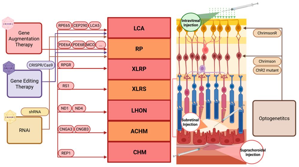 Figure 1. Application of AAV-mediated gene therapy in hereditary retinal diseases.
The Molecular Pathophysiology of Choroideremia
At the molecular level, Choroideremia is caused by a myriad of null mutations (deletions, nonsense, and frameshift mutations) in the CHM gene located on the X chromosome (Xq21.2). The CHM gene is responsible for encoding the Rab escort protein 1 (REP-1).
REP-1 is an indispensable component of the intracellular vesicular trafficking machinery. Its primary biological function is to bind newly synthesized Rab GTPases and present them to the enzyme Rab geranylgeranyl transferase (RabGGTase) for prenylation (the addition of geranylgeranyl lipid anchors). Following this post-translational modification, REP-1 escorts the prenylated Rab proteins to their specific target intracellular membranes.
In the absence of functional REP-1, a subset of Rab GTPases (particularly Rab27a and Rab38, which are crucial in the retina) remains unprenylated and inactive in the cytosol. In the retinal pigment epithelium (RPE) and photoreceptors, this disruption in vesicular transport leads to a catastrophic accumulation of undigested photoreceptor outer segments, defective melanosome transport, impaired phagocytosis, and ultimately, apoptotic cell death. Because CHM is a classic monogenic, loss-of-function disorder, it presents an ideal target for gene augmentation therapy.
Why Choroideremia Is an Ideal Target for Gene Therapy
Choroideremia presents a unique opportunity for therapeutic intervention:
-
Monogenic Disease
A single gene defect (CHM) simplifies therapeutic design and increases the likelihood of success. -
Localized Target Tissue
The retina allows for targeted delivery via subretinal injection. -
Slow Disease Progression
Offers a wide therapeutic window for intervention and monitoring. -
Established Gene Therapy Modality
AAV-based gene supplementation has shown promising safety and functional outcomes in clinical trials. -
Clear Biomarkers and Endpoints
Visual acuity, microperimetry, and imaging provide measurable clinical endpoints.
AAV Gene Therapy for Choroideremia
Choroideremia is a monogenic disease whose pathogenic gene CHM was successfully cloned in 1990. Thus, gene therapy is a preferred mean for choroideremia treatment. AAV vectors are a kind of tiny viruses, owning long-term protein expression through gene delivery, with high efficiency and non-toxic. Gene-enhancing therapy has been successfully validated in retinal disease models in dozens of animals by AAV vectors.
- AAV Vectors Design for Choroideremia
Human CHM cDNA was cloned into the transgene cassette of the AAV proviral plasmid, including the proximal chicken β actin (CBA) promoter and CBA intron 1 sequences flanking the CBA exon 1, to generate AAV8.CBA.hCHM. It also contains a 4.7 kb phage fragment fill, which can obtain AAV2/2 and AAV2/8 vectors by transfecting into HEK293T cells with the plasmid encoding the AAV2 or AAV8 vectors.
- AAV Vectors Research in the in Vitro Models for Choroideremia
To assess the ability of AAV8.hCHM to produce exogenous human REP-1 in vitro, it is necessary to analyze the expression and activity of its genes. The scientists found that a band of about 83 kDa of human REP-1 was appeared in the transfected cells, while no bands have appeared in the control group. Besides, western blotting was further used to characterize protein expression. A protein band of human REP-1 was clearly observed in transduced cell lysates, however, there were few bands in un-transduced cell lysates.
- AAV Vectors Research in the in Vivo Models for Choroideremia
Scientists indicate the efficacy of AAV treatment in CHMnull/WT mice via pupillometry and histology results. Although the mean amplitude of the eyes in the treated and control mice was similar (0.39 mm in the treated group and 0.34 mm in the control group), histology showed a marked improvement in retinal function in the treated group, which means that encoding hCHM into AAV vectors contributes to the recovery of choroidal function.
Key Challenges in Choroideremia Gene Therapy and Our Integrated Solutions
Developing effective gene therapies for choroideremia involves multiple technical and translational challenges. At Creative Biolabs, we address these hurdles through a combination of vector engineering, disease modeling, and advanced analytical platforms.
-
Efficient Transduction of Retinal Target Cells
Achieving robust transduction of both retinal pigment epithelium (RPE) and photoreceptors remains a key challenge.
Our Solution: We optimize AAV capsid selection (AAV2, AAV5, AAV8, engineered variants) and delivery strategies (subretinal vs. intravitreal) to maximize retinal coverage and transgene expression. -
AAV Packaging Capacity and CHM Cassette Design
The AAV packaging limit (~4.7 kb) requires careful design of the CHM expression cassette.
Our Solution: We design size-optimized constructs, including promoter selection, regulatory elements, and stuffer sequences, to ensure efficient packaging and stable expression. -
Sustained REP-1 Expression
Long-term therapeutic benefit depends on durable expression of functional REP-1 protein.
Our Solution: We screen promoter systems (CBA, CAG, CMV, retina-specific promoters) to balance expression strength and stability in retinal tissues.
Our Service Capabilities
Creative Biolabs provides comprehensive technical support spanning the entire gene therapy development pipeline for choroideremia:
| Services | Description |
|---|---|
| Vector Design & Optimization | Custom AAV vector construction with promoter selection (CBA, CMV, CAG, etc.), capsid engineering (AAV2, AAV5, AAV8, AAV9, and novel variants), and transgene optimization |
| In Vitro Model Validation | REP1 expression analysis (Western blot, immunofluorescence), functional prenylation assays, and cytotoxicity evaluation in CHM patient-derived fibroblasts and iPSC-RPE cells |
| In Vivo Model Development | Generation and characterization of CHM mouse models (Chmnull/WT), subretinal and intravitreal vector administration, and longitudinal disease monitoring |
| Pharmacodynamic Assessment | Pupillometry, electroretinography, optical coherence tomography, histology, and immunohistochemistry for therapeutic efficacy evaluation |
| Safety and Toxicology Studies | Comprehensive immunogenicity profiling, biodistribution analysis, and GLP toxicology support |
Comprehensive Analytical Services & Quality Control
Ensuring the quality, consistency, and functionality of AAV vectors is critical for successful gene therapy development. Creative Biolabs provides robust analytical and quality control services tailored to choroideremia programs.
Vector Characterization
We perform full characterization of AAV vectors, including genome integrity, capsid composition, and viral titer determination (qPCR, ddPCR).
Transgene Expression Analysis
REP-1 expression is evaluated using Western blot, immunofluorescence, and quantitative protein assays to confirm successful gene delivery.
Functional Assays
We conduct in vitro prenylation assays to verify the biological activity of REP-1 in restoring Rab GTPase function.
Retinal Imaging and Functional Evaluation
In vivo studies include optical coherence tomography (OCT), fundus imaging, electroretinography (ERG), and pupillometry.
Safety Assessment
We perform biodistribution studies, toxicity evaluation, and immunogenicity profiling to support preclinical development.
What Is the Ideal AAV Serotype for Choroideremia?
There is no single "universal" AAV serotype, but based on current research and clinical programs, AAV2 remains the gold standard, with several next-generation variants showing improved performance.
AAV Serotypes We Offer for Choroideremia Gene Therapy
| AAV Serotype | Retinal Tropism | Key Advantages | Limitations | Typical Use Case |
|---|---|---|---|---|
| AAV2 | Strong RPE, moderate photoreceptors | Clinically validated, high safety, widely used in CHM trials | Limited photoreceptor penetration | Primary choice for CHM gene augmentation |
| AAV5 | RPE + photoreceptors | Better spread than AAV2, good safety profile | Slightly lower clinical validation than AAV2 | Alternative for broader retinal coverage |
| AAV8 | Photoreceptors + RPE | High transduction efficiency, strong expression | Higher immunogenicity risk | High-expression CHM constructs |
| AAV9 | Broad retinal + systemic | Good tissue penetration | Less specific to retina | Exploratory or systemic approaches |
| AAV2/5 Hybrid | Enhanced retinal coverage | Combines AAV2 safety + AAV5 spread | More complex design | Optimized CHM delivery strategies |
| AAV2/8 Hybrid | Improved photoreceptor targeting | Strong expression + better penetration | Immunogenicity considerations | Advanced optimization programs |
| Engineered AAV (e.g., AAV2-7m8) | Enhanced photoreceptors (via intravitreal) | Superior retinal penetration, non-surgical delivery potential | Still emerging clinically | Next-gen CHM therapy development |
| Self-Complementary AAV (scAAV) | Same as parent serotype | Faster gene expression onset | Reduced packaging capacity | Small gene constructs like CHM (fits well) |
Why Choose Creative Biolabs as Your CHM Gene Therapy Partner?
In the highly competitive landscape of biopharmaceutical development, finding a reliable, scientifically rigorous CRO is paramount. Here is why top biotech firms and academic institutions choose Creative Biolabs for their inherited retinal disease pipelines:
- Deep Subject Matter Expertise: Our scientists possess decades of collective experience exclusively focused on AAV vectorology and ocular gene delivery. We understand the unique challenges of treating inherited retinal degenerations like Choroideremia.
- End-to-End Integration: Avoid the bottlenecks of transferring projects between multiple vendors. We offer a unified, streamlined pipeline from the initial in silico design of your CHM vector to the final in vivo safety report.
- Customization and Flexibility: We recognize that no two therapeutic programs are identical. We provide bespoke experimental designs, allowing you to choose the specific serotypes, promoters, cell lines, and animal models that best align with your strategic goals.
- Rapid Turnaround Times: In the race to the clinic, time is of the essence. Our optimized workflows and dedicated project management teams ensure that milestones are met promptly without ever compromising on data integrity or quality.
- State-of-the-Art Facilities: Our laboratories are equipped with the latest technologies in molecular biology, high-content imaging, animal housing, and viral manufacturing, ensuring that all data generated meets the highest regulatory standards.
What You Receive
Partnering with Creative Biolabs ensures that your choroideremia gene therapy program is supported with comprehensive deliverables at every stage.
- You will receive a fully customized AAV vector design strategy tailored to your CHM gene therapy objectives, including promoter selection, capsid optimization, and transgene configuration.
- Our team provides detailed plasmid maps and sequence documentation, ensuring transparency and reproducibility of your constructs.
- You will obtain high-quality AAV vectors with validated titers and full quality control reports, ready for downstream applications.
- For in vitro studies, we deliver complete datasets on REP-1 expression and functional restoration, including prenylation assay results and cellular validation.
- For in vivo programs, we provide comprehensive efficacy and safety evaluation reports, including imaging data, histological analysis, and functional assessments.
- In addition, you will receive a final project report summarizing experimental design, results interpretation, and recommendations for next-stage development, including IND-enabling strategies if required.
Frequently Asked Questions (FAQ)
Q: How does Creative Biolabs evaluate the functional rescue of REP-1 in vitro?
A: We do not merely confirm that the REP-1 protein is expressed (via Western Blotting, where hREP-1 appears as an 83 kDa band); we ensure it works. We perform specialized in vitro prenylation assays using protein lysates from transduced cells. By introducing unprenylated Rab proteins and radiolabeled geranylgeranyl pyrophosphate, we can quantitatively measure the ability of the AAV-delivered REP-1 to successfully facilitate Rab prenylation, which is the core biochemical deficit in Choroideremia.
Q: Can Creative Biolabs perform subretinal injections in small animal models?
A: Yes. Our highly trained veterinary surgeons possess the expertise to perform accurate subretinal injections in mice and other animal models, creating consistent blebs that cover a significant portion of the retina while minimizing surgical trauma.
Q: What size constraints exist when designing an AAV vector for the CHM gene?
A: AAV vectors have a strict packaging capacity of approximately 4.7 kilobases (kb). The human CHM cDNA is roughly 2.0 kb. This leaves ample room for regulatory elements (promoters, polyA signals, WPRE). In fact, the challenge is often avoiding a genome that is too small, which can lead to the packaging of truncated genomes or cellular DNA. To combat this, our vector engineering team meticulously designs "stuffer" sequences to optimize the cassette size closer to the ideal 4.7 kb, ensuring the production of high-quality, uniform viral particles.
Q: Which promoter is most suitable for CHM gene expression?
A: The choice of promoter depends on the target cell type and therapeutic goals. Ubiquitous promoters such as CBA and CAG are commonly used for robust expression, while retina-specific promoters can provide more targeted expression in RPE or photoreceptors. At Creative Biolabs, we evaluate multiple promoter options to identify the optimal balance between expression level, specificity, and long-term stability.
Q: What is the difference between subretinal and intravitreal delivery?
A: Subretinal injection delivers the vector directly to the RPE and photoreceptors, ensuring high transduction efficiency but requiring surgical expertise. Intravitreal injection is less invasive but may have limited penetration to outer retinal layers. We help clients select and optimize the most appropriate delivery route based on their research objectives.
Q: Can you support next-generation engineered AAV capsids?
A: Yes. In addition to standard serotypes such as AAV2, AAV5, AAV8, and AAV9, we also support engineered capsids (e.g., AAV2-7m8) with enhanced retinal penetration and transduction efficiency. These variants are particularly useful for improving photoreceptor targeting and enabling less invasive delivery strategies.
Get In Touch: Start Your CHM Gene Therapy Project Today
AAV vectors are widely used in the treatment of genetic diseases such as Choroideremia. Creative Biolabs provides comprehensive technical support, from vectors design to in vitro and in vivo model building and pharmacodynamic effect validation. Please contact us in time and we are glad to offer the highest quality service and guidance for you.
Reference
- Huang J, Li J, Xu X, et al. Adeno-associated virus vectors in retinal gene therapy: challenges, innovations, and future directions. Biomolecules, 2025, 15(7): 940. https://doi.org/10.3390/biom15070940 Distributed under Open Access license CC BY 4.0, without modification.