Product List Background CEA Aptamer Analysis
Background
Carcinoembryonic antigen (CEA) is a glycoprotein that participates in cell adhesion and is commonly found in body fluids, particularly serum. It is primarily produced by epithelial cells of the gastrointestinal tract during fetal development but has limited expression in normal adult tissues. Structurally, CEA belongs to the immunoglobulin superfamily, featuring multiple immunoglobulin-like domains. Increased levels of CEA expression are first detected in colon cancer, making it a valuable diagnostic marker for this malignancy. Elevated CEA levels are also found in other cancers, such as pancreatic, medullary thyroid, lung, and breast cancers, as biomarkers for disease progression and treatment response in these contexts.
Pathophysiology of CEA
CEA functions primarily in cell adhesion by binding to specific receptors on adjacent cells, facilitating intercellular interactions. It is detected in leukocytes, endothelial, and epithelial cells, promoting cell-cell adhesion through homophilic and heterophilic interaction with other subgroup proteins. Immunologically, CEA is classified as a member of the CD66 differentiation cluster, modulating immune responses. In tumors, elevated CEA levels contribute to tumor progression by enhancing cell aggregation, potentially aiding in metastasis by promoting tumor cell adhesion to vascular endothelium or distant organ surfaces. This adhesive property is crucial in the invasive behavior of cancer cells, impacting their ability to increase and spread within the body.
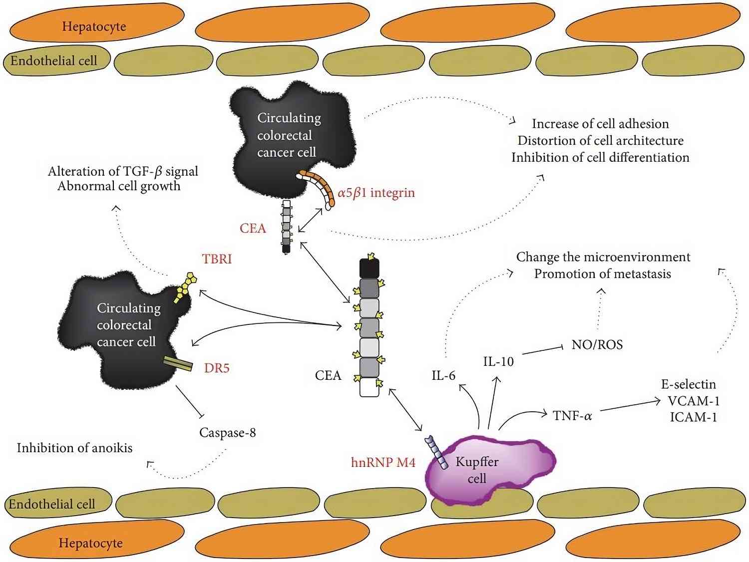 Fig.1 Schematic representation of CEA-affecting biological events.1,3
CEA in Colorectal Cancer Diagnosis and Post-Treatment Management
In colorectal cancer, CEA serves as a critical biomarker for diagnosis and post-treatment surveillance. A CEA level exceeding 5 µg/L at a new colorectal cancer diagnosis is linked to a poor prognosis. They are moreover, monitoring CEA levels in patients with colorectal cancer after initial therapy has proven helpful in detecting cancer recurrences. Rising CEA levels often precede clinical signs of recurrence, facilitating early suspicion and detection. So, CEA measurement should be performed every three months after surgery/treatment. Regular CEA testing helps clinicians assess the effectiveness of therapy and identify early signs of cancer recurrence. Early detection enhances the likelihood of a surgical cure through timely resectioning of recurrent tumors.
Diagnostic Significance of CEA in Other Cancers
Measurement of CEA levels is the most sensitive method for detecting surgically resectable liver metastases from colorectal cancer. In medullary thyroid cancer, CEA serves as a biomarker for disease progression and recurrence, aiding in monitoring therapeutic responses. In non-small cell lung cancer (NSCLC), greater CEA levels relate to advanced stage, nodal metastasis, and poor survival, influencing treatment strategies. In breast cancer, CEA is less commonly used but may indicate metastatic spread or recurrence in some cases. Overall, CEA's diagnostic utility across these cancers informs clinical decisions regarding treatment efficacy and disease management.
Creative Biolabs provides several aptamers targeting CEA with excellent specificity and affinity, allowing for accurate CEA detection in various situations. Our aptamers can help you speed up scientific research by providing quick reaction times and firm performance.
CEA Aptamer Analysis
Anti-CEA aptamers are synthetic oligonucleotides developed to bind specifically to CEA and are significant for early diagnosis, prognosis, and targeted cancer therapy. Aptamer selection technology, such as SELEX, allows for the generation of highly specific and strong binding affinity aptamers.
Anti-CEA aptamers function as recognition elements to be integral in advanced detection techniques like aptamer-based sensors and biosensors, providing rapid, sensitive detection of CEA in blood or tissue samples. These aptamers are invaluable for identifying tumor cells that overexpress CEA, offering a non-invasive method for cancer screening. They are used in targeted cancer diagnostics, allowing early identification of malignancies and monitoring of disease progression.
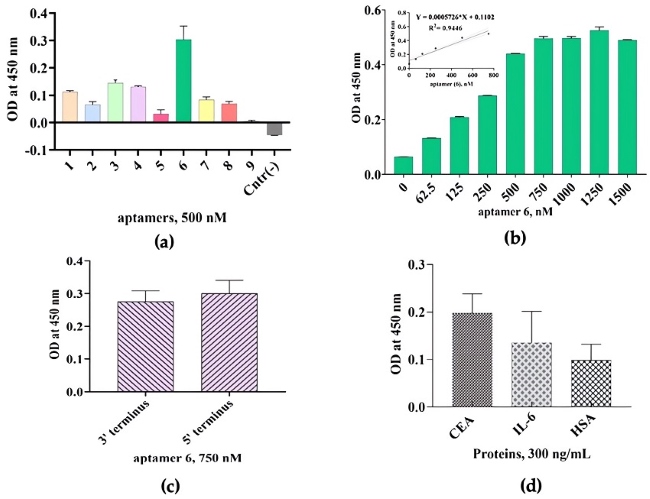 Fig.2 Binding capability and specificity study of the ssDNA aptamers towards CEA.2,3
Additionally, anti-CEA aptamers have demonstrated therapeutic potential, such as inhibiting liver metastasis of colon cancer cells by blocking CEA-mediated cell adhesion. They can also prevent cancer cell adhesion to endothelial cells, impeding tumor implantation and metastasis in vivo. This blocking ability represents a promising strategy for preventing cancer spread, improving the effectiveness of treatments, and reducing secondary tumor formation.
Creative Biolabs offers customized anti-CEA aptamer development, including high-affinity aptamer synthesis, assay optimization, and comprehensive support. Our services help customers achieve precise, rapid cancer detection and improved therapeutic outcomes in research projects, with a focus on personalized, targeted treatment strategies.
References
-
Lee, Joo Han, and Seong-Wook Lee. "The roles of carcinoembryonic antigen in liver metastasis and therapeutic approaches." Gastroenterology Research and Practice 2017.1 (2017): 7521987.
-
Yunussova, Nigara, et al. "A Novel ssDNA Aptamer Targeting Carcinoembryonic Antigen: Selection and Characterization." Biology 11.10 (2022): 1540.
-
Distributed under Open Access license CC BY 4.0, without modification.

 Datasheet
