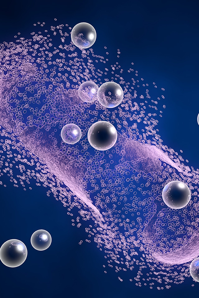

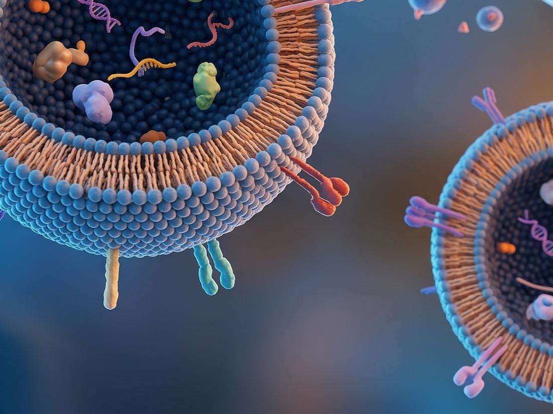
Integumentary system serves as a primary interface between the internal physiological environment and external stressors. Chronic exposure to ultraviolet radiation, chemical irritants, and environmental pollutants often precipitates inflammatory cascades, leading to manifestations such as erythema, pruritus, and compromised barrier integrity. In recent years, the paradigm of dermatological intervention has shifted toward biologics, with exosomes, emerging as pivotal mediators of intercellular communication. These lipid bilayers facilitate the horizontal transfer of proteins, microRNAs, and bioactive lipids, which modulate the skin microenvironment and promote homeostatic recovery.
Characterizing the soothing potential of exosome preparations is essential for translating basic research into therapeutic and cosmetic applications. Unlike conventional small molecule antiphlogistic agents, exosomes exert their effects through multifaceted signaling pathways, including the regulation of Toll-like receptors, the suppression of proinflammatory cytokine release, and the modulation of sensory neuron excitability. Accurate quantification of these effects is necessary to establish potency, safety, and mechanism of action. Rigorous efficacy testing ensures that candidate exosome products can mitigate skin sensitivity and accelerate the resolution of inflammation, providing a scientific foundation for product claims and regulatory compliance.
Engage Our Specialists to Refine Your Exosome Validation Protocol
Creative Biolabs offers a sophisticated suite of analytical services designed to evaluate the soothing and antiphlogistic properties of exosome samples. Our testing hierarchy spans molecular, cellular, and tissular assessments to provide a granular understanding of how cargoes derived from exosome influence skin health.
We examine the impact of exosomes on human keratinocytes by monitoring intracellular calcium flux and the expression of transient receptor potential vanilloid 1 (TRPV1) receptor. This service includes the quantification of nuclear factor kappa B (NF-κB) signaling pathway proteins and downstream inflammatory mediators such as endothelin and various interleukins.
Utilizing RAW 264.7 or primary macrophage models, we assess the capacity of exosomes to suppress inflammation induced from lipopolysaccharide. We measure changes in the secretion of tumor necrosis factor alpha (TNFα), IL6, and cyclooxygenase 2, alongside the upregulation of antiphlogistic markers.
To simulate complex tissue interactions, we employ 3D reconstructed human epidermis (RHE) models. This allows for the evaluation of exosome penetration and its subsequent effect on the suppression of chemically induced erythema and the modulation of the inflammatory secretome within a stratified tissue structure.
Our scientists analyze the influence of exosomes on mast cell degranulation. By observing morphological changes and quantifying the release of histamine and proteases, we evaluate the potential of the sample to reduce immediate hypersensitivity reactions.
We utilize Chinese Hamster Ovary (CHO) cells engineered to express specific histamine receptors. This system measures the inhibitory rate of exosome samples on receptor activity, providing data on the antipruritic potential of the vesicles.
Given the link between skin sensitivity and the nervous system, we conduct assays using neuronal cell lines to monitor calcium signaling, TRPV1 expression, and the release of neuropeptides such as Substance P.
Consult With Our Biologists to Align Testing with Your Product Goals
The integration of exosomes with documented soothing efficacy into product pipelines offers significant strategic benefits for developers.
Exosomes facilitate the efficient delivery of bioactive molecules across the stratum corneum, often surpassing the penetration depth and stability of free synthetic compounds.
These vesicles typically exhibit lower cytotoxicity and a reduced risk of skin sensitization compared to traditional chemical actives, aligning with the growing consumer demand for biological ingredients.
Developers can create products that address multiple pathways simultaneously, such as combined antiaging and antiphlogistic benefits, by utilizing exosome preparations with characterized proteomic and transcriptomic profiles.
Rigorous data generation during the development phase streamlines the safety assessment process and ensures compliance with international standards for cosmetic and dermatological ingredients.
Discuss Your Project Requirements with Our Exosome Research Team
The scientific validity of skin soothing mediated by exosome is supported by a growing body of literature. Recent investigations have highlighted the diverse origins of these vesicles and their unique functional signatures. For instance, exosomes derived from bovine milk have demonstrated significant antiphlogistic activity. Research indicates that these vesicles can promote the reconstruction of stratum corneum lipids and enhance barrier function. In macrophage models, these bovine milk exosomes attenuate the secretion of proinflammatory cytokines such as IL6 and TNFα while simultaneously increasing the expression of antiphlogistic factors. These findings suggest that milk exosomes act as potent soothing agents by reprogramming the immune response.
Similarly, exosomes derived from plant, such as those isolated from apple, have shown efficacy in vitro settings. Studies involving vesicles derived from apple (ADVs) indicate a marked reduction in skin redness induced by chemical. These vesicles appear to operate through a sustained release mechanism, providing prolonged relief from irritation without the side effects associated with topical corticosteroids. The success of ADVs in reducing wrinkle volume and skin roughness also points to a dual action role where soothing effects complement antiaging pathways. Our testing services are modeled after these high impact studies, ensuring that the parameters we measure are those most relevant to physiological outcomes.
Connect With Our Scientists to Optimize Your Efficacy Evaluation Plan
Creative Biolabs operates a specialized exosome engineering platform that provides a solution for the entire developmental lifecycle:
 Precision Isolation and Purification
Utilizing advanced techniques such as tangential flow filtration (TFF), ultracentrifugation, and size exclusion chromatography to ensure high yield and purity of intact vesicles.
Rigorous Characterization
Validation of exosome identity through nanoparticle tracking analysis (NTA) for size distribution, transmission electron microscopy (TEM) for morphology, and western blotting for specific surface markers like CD63 and CD81.
Functional Cargo Engineering
Capabilities to load exosomes with specific therapeutic payloads or modify surface proteins to enhance targeting to specific dermal or epidermal cells.
Standardized In Vitro Assays
Integration of the soothing efficacy tests described above with standardized protocols to ensure reproducibility and scientific rigor across different batches.
Customized Pilot Production
Support for scaling up the production of characterized exosome samples for further testing or integration into final formulations.
Request a Technical Session to Secure Your Efficacy Testing Strategy
A: Our laboratory is equipped to handle vesicles from various origins, including mesenchymal stem cells (MSCs), plant (fruits, vegetables, or herbs), and bovine milk. We adapt our protocols to account for the customer needs.
A: Prior to conducting efficacy assays, we perform cytotoxicity evaluations such as CCK8 or MTT assays. This ensures that any observed reduction in inflammatory markers is due to the bioactive properties of the exosomes rather than a decrease in cell viability.
A: Clients receive a technical report containing raw data, statistical analysis, high resolution imaging (where applicable), and a professional interpretation of the findings relative to industry standards and control groups.
A: Our platform includes dose and response studies as a core component of the testing service. We help identify the minimum effective concentration required to achieve the desired soothing effect, which is critical for formulating final products.
Speak With Our Lead Researchers Regarding Your Exosome Sample Analysis