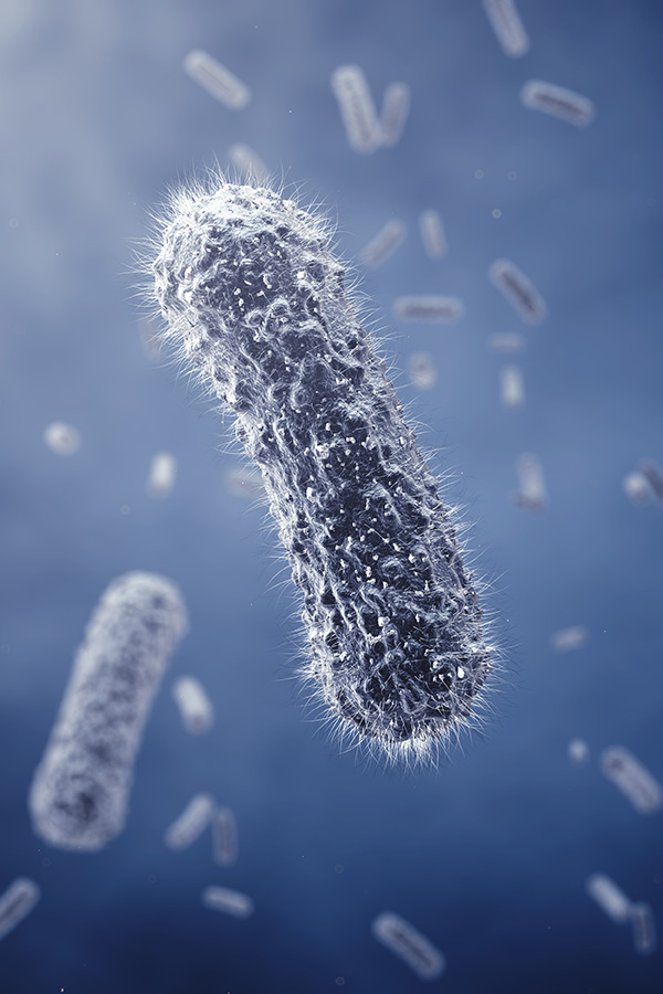

Helicobacter pylori (H. pylori) is a microaerophilic, Gram negative bacterium that has evolved to thrive within the hostile, acidic environment of the human stomach. Infecting approximately half of the global population, this pathogen is the primary etiological agent for chronic gastritis, peptic ulcers, and gastric adenocarcinoma. While conventional microbiology once focused predominantly on direct cell to cell contact and the secretion of soluble toxins, the paradigm has shifted toward the role of exosome.
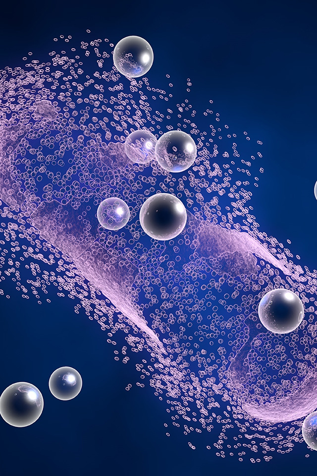
In the broader biomedical landscape, these bacterial vesicles are often discussed alongside mammalian exosomes due to their similar nanoscale dimensions and lipid bilayer architecture. H. pylori constitutively shed these biogenic nanocarriers, which act as sophisticated delivery proteoliposomes. They encapsulate a concentrated repertoire of virulence factors, lipids, and small RNAs, shielding them from degradation by extracellular enzymes and gastric acid. Investigating H. pylori derived vesicles is essential not only for deciphering gastric pathogenesis but also for understanding how this localized infection exerts systemic effects. Recent evidence connects these vesicles to extra gastric pathologies, including neurological impairment, vascular inflammation, and hepatic fibrosis.
The biological utility of H. pylori vesicles stems from their unique structural and functional attributes. These vesicles provide the bacteria with a multifaceted tool for environmental adaptation and host manipulation.
| Feature | Pathobiological and Technical Utility |
|---|---|
| Protective Encapsulation | The rigid bilayer protects labile cargo such as mRNA and fragile proteins from the proteolytic environment of the gut. |
| Systemic Translocation | Their small size allows them to penetrate the gastric epithelial barrier and enter the bloodstream, reaching the blood brain barrier or liver. |
| High Local Concentration | Vesicles deliver a "package" of multiple toxins simultaneously to a single target cell, triggering a more potent response than soluble monomers. |
| Surface Mimicry | The presence of Lewis antigens and adhesins on the vesicle surface enables highly specific binding to host receptors like CD44 or TLRs. |
| Non Replicative Safety | As acellular entities, they retain the immunogenicity of the parent strain without the risks associated with live bacterial infection. |
Consult With Our Team to Utilize These Distinctive Traits in Your Experimental Design.
The study of H. pylori derived nanocarriers has expanded from basic morphological characterization to deep mechanistic inquiries into host pathogen crosstalk. These vesicles are now recognized as central players in both local and systemic disease progression.
Vesicles from H. pylori carry upwards of 400 distinct proteins that induce radical proteomic shifts in gastric epithelial cells. Upon internalization, they activate the PI3K-Akt pathway and stimulate NFκB, leading to a massive release of CXCL8 and other proinflammatory cytokines. This chronic inflammatory state is a precursor to atrophic gastritis and eventual malignant transformation.
A groundbreaking area of research involves the translocation of these vesicles to the Central Nervous System (CNS). In murine models, orally administered vesicles have been detected in the brain parenchyma. Once they cross the biological barriers, they are predominantly taken up by astrocytes. This leads to:
Beyond the brain, these nanocarriers contribute to atherosclerosis by impairing the proliferation of vascular endothelial cells and inducing oxidative stress. In the liver, they have been observed to activate hepatic stellate cells, increasing the expression of fibrosis markers and inhibiting healthy autophagy flows.
Align With Our Analytical Leads to Streamline Your Investigation into These Systemic Pathways.
The intrinsic characteristics of H. pylori vesicles render them exceptional candidates for conversion from laboratory subjects to therapeutic entities. Their capacity to regulate the immune system and focus on particular tissues presents numerous translational opportunities.



Talk To Our Experts About How to Turn These Bacteria Carriers into Helpful Tools.
Creative Biolabs offers a range of services that help researchers get past the technical problems that come up when studying microbial vesicles. Our infrastructure supports every step of the development pipeline, making sure that the data is clean and can be reproduced.


Talk To Our Technical Leads Today About the Goals of Your Experiment.
Recognizing the need for standardized materials in complex microbiological studies, we offer tailor made vesicle preparations. These products are manufactured under stringent quality control to ensure consistency across different batches.
Vesicles pre labeled with bioluminescent or fluorescent markers for real time tracking in cellular or animal models.
Custom loading of exogenous molecules, such as siRNA, small molecule inhibitors, or specific proteins, into the vesicle lumen for delivery research.
Every preparation undergoes rigorous 0.22 μm filtration and plating to ensure the total absence of viable bacteria.
Collaborate With Us to Engineer Your Specialized Delivery Systems.
A: Studies show that they can move through the bloodstream after crossing the damaged gastric mucosa. Because they are small and made of lipids, they can interact with and eventually get past the blood-brain barrier, probably through transcytosis or by taking advantage of areas that are more permeable during systemic inflammation.
A: The structural integrity of the lipid bilayer is our top priority. We usually ship our vesicles in special buffers at very low temperatures (-80 °C) to keep the cargo from leaking and the membranes from fusing. This makes sure that they arrive ready for functional tests.
A: Yes. It is known that bacterial vesicles carry resistance determinants, such as β-lactamase enzymes. They can be used to learn about horizontal gene transfer and how pathogens protect themselves from antimicrobial pressure.
A: Exosomes come from the outer membrane of Gram-negative bacteria and have bacterial-specific markers like LPS and peptidoglycan. Exosomes, on the other hand, come from the endosomal pathway of eukaryotic cells.
Get In Touch with Our Scientists to Start Your Next Discovery.