GTOnco™ Phagocytosis Assay Services
Phagocytosis is a complex process for the ingestion and elimination of pathogens, and also participates in the elimination of apoptotic cells. The specialized immune cells including macrophages, neutrophils, and dendritic cells have phagocytic functions and also participate in specific immunity, playing an important role in cellular immunity. Creative Biolabs manifests as a world-leading expert in I-O drug discovery field and provides our clients with top-quality phagocytosis assays. Our phagocytosis assay services can be designed to analyze the function of specialized immune cells and evaluate the anti-tumor activity of gene therapy-based I-O products.
Phagocytosis Assay Introduction
Phagocytosis is a complex process involving a series of steps: identifying target particles, activating signaling pathways for internalization mechanisms, forming phagosomes, and maturing them. Phagocytosis plays an important role in anti-infection. Phagocytosis assay is an important in vitro tool for quantifying the uptake of particles (such as bacteria, apoptotic cells, regulatory targets) by specialized phagocytic cells (mainly macrophages, neutrophils, and dendritic cells (DCs)). The quantitative measurement of phagocytic activity using this assay is crucial for evaluating the efficacy of therapeutic antibodies, such as those acting through antibody dependent cellular phagocytosis (ADCP), and understanding the immune evasion mechanisms employed by pathogens or tumor cells. Its relevance covers oncology, infectious diseases, autoimmune and inflammation research.
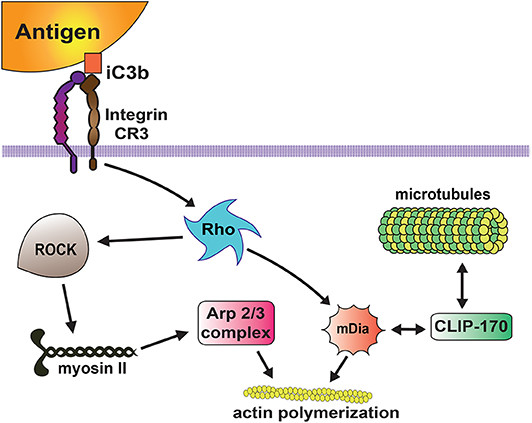 Figure 1 Complement receptor signaling for phagocytosis.1
Phagocytosis Mechanism
Phagocytosis is a three-step cellular process for target clearance:
- Recognition and Binding: Phagocytes use PRRs (Pattern Recognition Receptors) to detect targets. Opsonization, where targets are coated with host proteins like antibodies, significantly enhances uptake via Fc receptors, a principle central to ADCP.
- Internalization: Receptor binding triggers actin cytoskeleton rearrangement, leading to the formation of pseudopods that engulf the target, forming a membrane-bound vesicle called the phagosome.
- Killing and Degradation: The phagosome fuses with the lysosome, forming an acidic. Targets are destroyed by Reactive Oxygen Species (ROS), generated by NADPH oxidase, and lytic enzymes.
Key Signaling Pathways in Phagocytosis
| Receptor Type | Signaling Pathway | Functional Outcome |
|---|---|---|
| Toll-like Receptors (TLRs) | Actin-Cdc42/Rac, p38 MAPK | Pathogen recognition and clearance |
| Fc Receptors | SYK, PI3K, RAC1 | Antibody-dependent phagocytosis |
| Complement Receptors | RHOA, RAC1 | Complement-mediated particle internalization |
| Scavenger Receptors | Various adaptor proteins | Cellular debris clearance |
Phagocytosis Assay Flow Cytometry for Macrophages for Mouse
Flow cytometry is the gold standard for highly quantitative and multiplex phagocytic detection, providing single-cell resolution. For mouse macrophages, this method can accurately characterize subpopulations of phagocytic cells.
- Macrophage Isolation: Harvest mouse bone marrow-derived macrophages (BMDMs) or peritoneal macrophages. For BMDMs, culture bone marrow cells from C57BL/6 mice in DMEM + 10% FBS + 20 ng/mL M-CSF for 7 days to induce differentiation.
- Target Labeling: Use fluorescently labeled targets.
- Phagocytosis Incubation: Seed 1×10⁵ macrophages/well in 96-well plates. Add labeled targets at a target-to-phagocyte ratio (T:P) of 10:1 (optimized for BMDMs) and incubate at 37°C for 1–2 hours (4°C control to block active phagocytosis).
- Flow Cytometry Analysis: Wash cells to remove unbound targets, stain with macrophage markers (e.g., CD11b-PE, F4/80-APC), and analyze on a flow cytometer.
Types of Phagocytosis Assay
Neutrophil Phagocytosis Assay
Focus: It is crucial for acute bacterial infections and measuring the bactericidal ability of neutrophils, which are the most abundant and rapidly recruited phagocytic cells.
Method: Regularly use conditioning bacteria or latex beads. Flow cytometry is common, but microscopy is also highly effective for morphology-based analysis. ROS generation (detected during oxidation using dyes such as DHR 123) is usually measured simultaneously as a functional reading.
Macrophage Phagocytosis Assay
Focus: It is crucial for chronic inflammation, ADCP research (such as therapeutic antibodies in oncology), and infectious cell diseases (clearing apoptotic cells).
Method: Highly versatile; Perform high-throughput screening (HTS) using flow cytometry, live cell imaging, or ELISA based formats. The target range ranges from CFSE labeled tumor cells to IgG regulated red blood cells (RBCs) in classical phagocytic index assays.
Dendritic Cells Phagocytosis Assay
Focus: Antigen presentation and bridging innate and adaptive immunity. DC phagocytosis is used to obtain antigens for processing and presentation to T cells.
Method: Typically, uptake of low abundance targets such as peptides, viruses, or protein coated beads is measured. Due to its lower phagocytic ability compared to macrophages/neutrophils, flow cytometry requires high sensitivity. The downstream steps of MHC class I/II presentation are usually evaluated together with uptake.
Applications of Phagocytosis Assays
Phagocytosis test is indispensable in modern biomedical research:
- Immunotherapy development (oncology): Measure the ADCP of Fc engineered monoclonal antibodies (e.g., determine the quality of next-generation anti-CD20 or anti-HER2 antibodies).
- Infectious diseases: Assess the ability of host phagocytes to clear new or drug-resistant pathogens and screen for new candidate vaccines that depend on regulatory antibodies.
- Inflammation and Autoimmunity: Investigate the dysregulation and clearance of disease-associated immune complexes or apoptotic fragments (e.g., systemic lupus erythematosus SLE).
- Drug safety/efficacy: Evaluate the impact of small molecules on phagocytic cell function.
Core Services at Creative Biolabs
At Creative Biolabs, we offer a comprehensive portfolio of at GTOnco™ phagocytic assay services designed to meet the diverse needs of our clients in academic research, drug development, and clinical diagnostics. Our services leverage decades of expertise in immunology and cell biology, combined with state-of-the-art instruments and strict quality control measures.
Our service portfolio includes:
- Customizable at GTOnco™ phagocytic assays of macrophages, neutrophils, and dendritic cells from human, mouse, and other model species
- High content screening platform utilizing recombinant target particles with specific membrane composition
- Multi parameter flow cytometry analysis capable of simultaneously evaluating phagocytosis, oxidative burst, and cell surface markers
- Real time imaging analysis of mature living cells using pH sensitive probes for monitoring phagosome maturation
- Specialized detection methods for evaluating phagocytic acidification, antigen treatment, and bactericidal activity
Preparation and Labeling of Phagocytic Targets
Common targets include heat inactivated microorganisms such as Staphylococcus aureus and Escherichia coli, fluorescent synthetic beads, and apoptotic cells. Each target type has its unique advantages: microorganisms provide physiological relevance, beads provide uniformity and bright fluorescence, apoptotic cells can study efferent cell diseases (clearing dying cells). At Creative Biolabs, we have developed a complex target preparation scheme using recombinant membrane coated particles combined with specific molecular components to study specific phagocytic pathways.
Table 1 Comparison of Phagocytic Target Labeling Strategies
| Labeling Method | Advantages | Limitations | Applications |
|---|---|---|---|
| CFSE | Stable intracellular fluorescence, cell tracing compatible | Potential cytotoxicity at high concentrations | Long-term tracking, flow cytometry |
| SYTOX Green | pH-independent fluorescence, nucleic acid binding | Requires permeabilized targets | Bacterial phagocytosis quantification |
| FITC | Bright fluorescence, well-established protocols | pH-sensitive, fluorescence quenched in acidic phagosomes | General phagocytosis assays |
| Reconstituted Membranes | Controlled composition, customizable ligands | Technical complexity, specialized expertise required | Mechanism studies, receptor-ligand interactions |
Our Methods
Our solution has been validated and meets strict standards at GTOnco™ platform, ensuring high-quality and reproducible data suitable for regulatory submission and publication.
High-Content/High-Throughput Flow Cytometry
utilizing advanced cell sorting instruments and analyzers for multi parameter analysis (phagocytosis, survival rate, activation markers).
Live-Cell Confocal Microscopy
provides real-time 3D tracking of target uptake and phagosome maturation, particularly suitable for studying dynamics and mechanism details.
ELISA/Colorimetric Assays
used for high-throughput preliminary screening of large-scale compound libraries, typically using biotinylated targets captured by reporter beads or phagocytic cells.
Features of Our Phagocytosis Assays at GTOnco™ Platform
- Custom assay services and advanced analysis techniques are available;
- Various types of specialized immune cells we can handle: macrophages, neutrophils, and dendritic cells;
- Fast turnaround time and superior quality.
Frequently Asked Questions
Q: How do you distinguish between surface bonding and true internalization?
A: We mainly use two gold standard methods:
- Fluorescence quenching: The application of chemical quenchers (such as trypan blue) or specific antibody-based quenchers to eliminate signals from non-internalized, surface bound fluorescent targets.
- PH sensitive infectious material: Using probes such as pHrodo, high fluorescence is only emitted in acidic pH environments where lysosomes are engulfed, ensuring that only successfully internalized and trafficked targets are measured.
Q: Which phagocytic assay is most suitable for studying antigen presentation?
A: For research focused on antigen presentation, dendritic cell phagocytosis assay is the most suitable, as dendritic cells specialize in antigen preservation and presentation. In addition to phagocytic uptake, these tests typically also include measurements of MHC expression and T cell activation.
Q: How do you distinguish between truly internalized targets and targets that only adhere to the cell surface?
A: We employed several techniques for this critical differentiation, including trypan blue quenching of extracellular fluorescence, differential fluorescence labeling of internalized particles and surface bound particles, and confocal microscopy with z-slices to verify intracellular localization.
Q: What controls are included in your phagocytic test?
A: Our detection method includes appropriate positive controls (e.g. targets regulated with known ligands) and negative controls (e.g. targets incubated at 4 ° C to inhibit phagocytosis, untreated with ultrasound). For inhibitor research, we include carrier controls and, where possible, validate the specificity of inhibitors.
Connect with Us Anytime!
Taking advantage of the GTOnco™ platform, Creative Biolabs is able to conduct many in vitro tests to evaluate the phagocytic functions and therapeutic efficacy of gene therapy-based I-O drugs. we will apply the rigorous criteria to generate reliable and solid data for every research project. For more detailed information, please feel free to contact us.
Reference
- Uribe-Querol E, Rosales C. Phagocytosis: our current understanding of a universal biological process. Frontiers in immunology, 2020, 11: 1066. https://doi.org/10.3389/fimmu.2020.01066 (Distributed under Open Access license CC BY 4.0, without modification.)