K1L-based Tumor Selectivity Enhancement of Oncolytic Vaccinia Virus
With the exploration of oncolytic vaccinia virus (VACV) development for many years, Creative Biolabs has established an advanced VACV engineering platform, transforming VACV to meet customers' requirements in research, preclinical study, and drug development.
VACV is a kind of genetically engineered or naturally occurring virus, which can selectively replicate in and kill cancer cells without damage to the normal tissues. VACV is usually highly pathogenic and may cause damage to humans. Therefore, the design and manipulation of the viral genome to produce a non-pathogenic virus have become a standard method for VACV development. Nowadays, VACV therapy has become a potential new option for treating cancer.
Scientists from Creative Biolabs provide a full range of pathogenicity manipulation methods for the development of the vaccinia virus (VV). We provide customized solutions for our clients according to your projects.
Brief Introduction of K1L Gene
The VV K1 protein is a 31 kDa intracellular protein encoded by the K1L gene. K1L was initially described as a host range factor because its expression is necessary for VV replication in rabbit and hamster cell lines. K1L is an important gene regulating the host range of the VV, including the infectivity of the VV in human cells. One copy of K1L is enough to make the VV overcome the replication restriction in pig kidney cells. It is now known that K1 has other functions of antagonizing immune response. K1 can antagonize NF-kβ, PKR, IRF1 and IFN signal transduction pathways.
VV with Deletion or Mutation in the K1L Gene
- Decrease VV Pathogenicity
- Decrease VV Replication In Vivo
- Modulation of Innate Immunity
The pathogenicity of the VV is usually decreased by mutation of the K1L gene. Scientists have reported that a virus lacking the K1L gene is less pathogenic than its wild-type parental. In addition, the reinsertion of K1L into a VV that has removed the K1L gene (vΔK1L) has caused pathogenicity similar to that of wild-type VV (vK1L), suggesting that the K1L gene is responsible for the decrease of pathogenicity.
Usually, a reduction in virus pathogenicity is associated with the decrease of virus replication in mouse organs. For example, vΔK1L titer in the lungs is approximately 10 times lower than the vK1L on day 2, and 100 times lower on days 4 and 6.
VV infection with the deletion of K1L raised the expression of TNF in cell culture. The decrease of immune response gene expression was associated with the decrease of leukocyte infiltration and smaller lesion size.
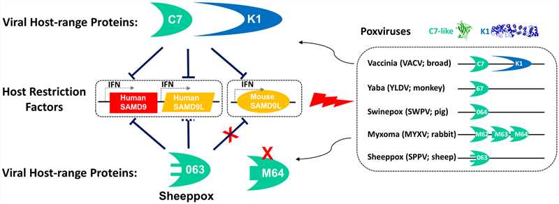 Fig.1 Summary of SAMD9/L restriction factors in human and mice and their antagonism by different poxviruses. (Meng, 2018)
The K1L deleted VV may be a valuable tool for further understanding the relationship between viral pathogenesis, the regulation of innate immunity, and the stimulation of protective immune response.
With our mature VACV engineering platform, the experienced scientists at Creative Biolabs are committed to helping you develop a unique VACV. We also provide other various services regarding VACV development.
Reference
- Meng, X.; et al. A paralogous pair of mammalian host restriction factors form a critical host barrier against poxvirus infection. Plos Pathogens. 2018, 14(2).