Reporter-encoding Oncolytic Virus
Oncolytic viral therapies now become a potential therapeutic option in the cancer research world due to the increased treatment efficacy and reduced side effects. Pre-clinical studies need to noninvasively and serially identify sites of viral targeting via molecular imaging in order to provide efficacy, safety and toxicity information. In addition, molecular imaging of oncolytic viral therapy has contributed to detecting tumor origin and presence of metastases sensitively and specifically. Molecular imaging can improve oncolytic virus vector design and clinical protocols for future personalized treatments with the information of the useful viral dose and administration schedule. Creative Biolabs provides multiple strategies for molecular imaging of oncolytic virus replication and several imaging methods including photoacoustic imaging, computer tomography (CT), magnetic resonance imaging (MRI), single photon emission CT (SPECT), positron emission tomography (PET) and gamma-scintigraphy.
Optical Reporter Gene Systems
Oncolytic viruses encoding reporter genes utilized for in vivo molecular imaging are useful to locate the distribution of oncolytic viruses in pre-clinical tests. Optical detection methods mainly include green fluorescent protein (GFP), enhanced GFP (eGFP), discosoma red fluorescent protein (DsRed), and bioluminescence imaging (BLI), which utilizes luciferases. Reporter-encoding oncolytic viruses, including vaccinia virus, adenovirus, herpes simplex virus and vesicular stomatitis virus, allow accurate tracking of gene expression, tumor metastases, viral infection as well as assessment of gene therapy.
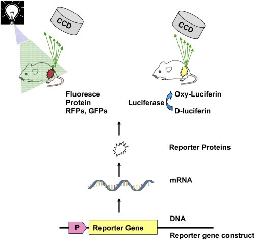 Fig 1. Monitoring gene expression using optical imaging.
Advantages of optical detection methods
- Short acquisition times
- High spatial resolution
- Higher sensitivity
- Lower background luminescence
Deep-tissue Reporter Gene Systems
Four types of deep-tissue reporter genes have been used for oncolytic viral therapies.
- The enzyme reporter gene: herpes simplex virus-thymidine kinase (HSV1-tk) encoded by vaccinia virus, adenovirus and herpes simplex virus.
- Human norepinephrin transporter (hNET) / Human sodium iodide symporter (hNIS) reporter gene: encoded by vaccinia virus, vesicular stomatitis virus, measles virus, adenovirus and herpes simplex virus.
- Human somatostatin receptor 2 (hSSRT2) reporter gene: encoded by vaccinia virus.
- Contrast agents: melanin encoded by vaccinia virus.
Advantages of Deep-tissue detection methods
- Allowing weak promoters and low levels of gene expression
- Repeatability, quantifiability and high sensitivity
Creative Biolabs provides a series of purified reporter-encoding oncolytic virus, including herpes simplex virus and vaccinia virus. Except for the following existing products, we provide customized services to meet the specific need of our clients for different reporter genes.
Reference:
- Youn, H., & Hong, K. J. (2012). In vivo noninvasive small animal molecular imaging. Osong public health and research perspectives, 3(1), 48-59.