Vaccinia Growth Factor-based Tumor Selectivity Enhancement of Oncolytic Vaccinia Virus
Overview of Vaccinia Growth Factor
The inverted terminal repetition (ITR) of the vaccinia virus genome contains at least three genes that are expressed early during infection. A portion of the predicted amino acid sequence of one has been shown by computer analysis to be closely related to epidermal growth factor (EGF) and transforming growth factor-alpha (TGF-α) and more distantly related to proteins of the blood coagulation system. Analysis of vaccinia virus-infected culture supernatants led to the detection of an acid-stable polypeptide that competed with EGF for binding to EGF membrane receptors, induced phosphorylation of tyrosine residues in the EGF receptor, had mitogenic properties for tissue culture cells, did not cross-react with TGF-α in a radioimmunoassay, and exhibited minimal reactivity with certain antisera for native EGF. Since this EGF-like activity was present only in virus-infected cultures and was synthesized with the kinetics expected of an early virus-encoded gene product, it was termed vaccinia virus growth factor (VGF).
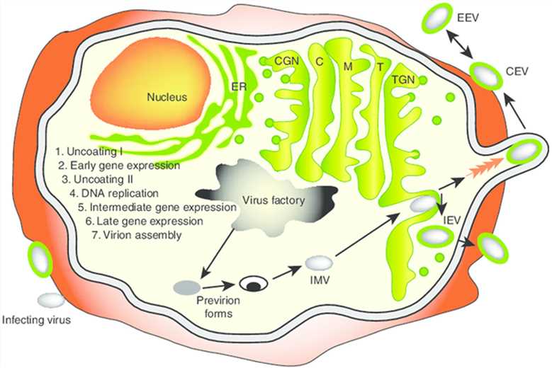 Fig.1 Vaccinia virus life cycle. (Guo, 2019)
Researches for the Deletion of VGF
A VGF- virus was constructed by removing approximately half of both viruses' VGF genes and replacing them with a cassette expressing a color selection marker, i-galactosidase. It was shown that this mutant no longer produced a factor(s) that competes with EGF for binding to the EGF receptor, stimulated the anchorage-independent growth of appropriate rodent cells in the presence of TGF-α, or triggered phosphorylation of tyrosine residues in the EGF receptor. From these observations, it can be inferred that no other region of the vaccinia virus genome codes for a protein capable of specific binding to EGF receptors.
Besides, the insertion of the P-galactosidase cassette into the virus genome of the VGF- mutant did not affect virus replication under optimal cell growth conditions in BS-C-1 cells. According to a number of criteria, intradermal inoculations of the rabbit with WT virus resulted in lesions that were more severe than those caused by the VGF- mutant. It was further observed that at almost all doses of virus, the WT virus appeared to induce localized cellular hyperplasia at the advancing margins of the lesion, whereas the mutant did not. This cellular hyperplasia could have resulted from the direct action of VGF synthesized in infected cells or, alternatively, from the host-encoded growth factors released from infected cells or as part of the wound-healing process.
Analysis of virulence genes such as VGF has taken on renewed importance with the potential application of the vaccinia virus as a universal vaccine vehicle. For the safe use of these recombinant vaccinia virus vaccines, the virus virulence may need attenuation to reduce the risks of post-vaccinial complications. The VGF- mutation may be a desirable attenuation marker for inclusion in the vaccinia virus recombinant vaccine strains.
Services at Creative Biolabs
Struggling in the field of oncolytic virus enhancement over years, Creative Biolabs has gradually optimized our platform with advanced facilities, the latest technologies, and excellent experts specialized in this field. Strong foundations are our reliable guarantee for offering customer-satisfied enhancing tumor selectivity services based on the vaccinia growth factor to global clients. Every effort we make is dedicated to providing better service to our customers.
Reference
- Guo, Z. S.;et al. Vaccinia virus-mediated cancer immunotherapy: cancer vaccines and oncolytics. J Immunother Cancer. 2019, 7(1): 6. Distributed under Open Access license CC BY 4.0, without modification.