GTOnco™ Macrophage Polarization Assay Services
Macrophages exert multiple functions including modulating the adaptive immune responses and responding to pathogens. Macrophages show specific functions and phenotypes in response to different stimuli. They can display polarized states via their plasticity and different populations of macrophages can be developed in response to different stimuli. The macrophage polarization has been classified as being either M1 (pro-inflammatory) or M2 (anti-inflammatory) state, which is further divided into M2a, M2b, M2c and M2d subsets. At Creative Biolabs, we offer custom assays to induce different macrophage subsets and characterize their polarization states.
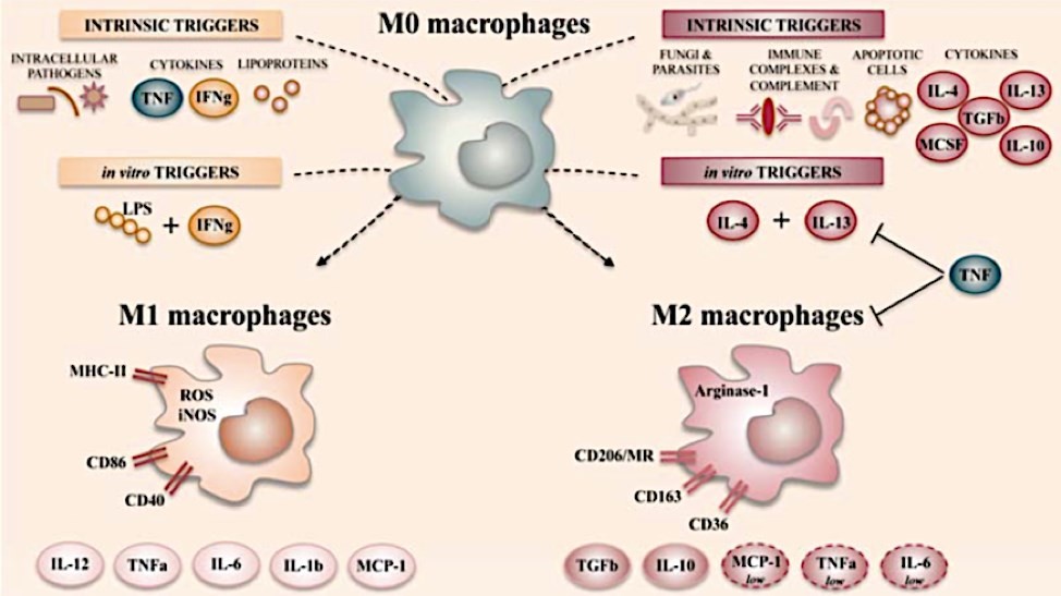 Figure 1 The main polarization states of activated macrophages.1
Macrophage Polarization Assay
Macrophage polarization represents a spectrum of activation states, generally divided into two major extremes: the pro-inflammatory, microbicidal M1 phenotype (classical activation) and the pro-resolving, tissue-remodeling M2 phenotype (alternative activation), with additional M2 subtypes (M2a, M2b, M2c, M2d).
Table 1 Macrophage Polarization Assay Phenotypes.
| Phenotype | Key Inducers | Primary Functions |
|---|---|---|
| M1 | IFN-γ, LPS | Host defense, Th1 promotion, cytotoxicity, increased antigen presentation |
| M2 | IL-4, IL-13 (M2a); Immune Complexes, TLR ligands (M2b); IL-10, TGF-β (M2c) | Tissue repair, Th2 promotion, resolution of inflammation, tumor promotion/angiogenesis |
Macrophage Polarization in Vitro Assay
In vitro polarization assays provide a controlled system to dissect the complex signaling and metabolic pathways that control macrophage function, unaffected by the confounding variables of the in vivo environment. The selection of starting cell materials is a critical first step, and each model system offers unique advantages.
Table 2 Common Cell Models for In Vitro Macrophage Polarization Assays.
| Cell Model | Description | Key Polarization Stimuli | Advantages | Considerations |
|---|---|---|---|---|
| Primary Human Monocytes | Isolated from peripheral blood mononuclear cells (PBMCs) of donors. |
M1: GM-CSF, followed by LPS + IFN-γ M2: M-CSF, followed by IL-4 + IL-13 |
Most physiologically relevant, human-specific immune responses. | Donor variability, limited expansion capacity, requires ethical approval for collection. |
| THP-1 Cell Line | Human monocytic leukemia cell line. |
Differentiation with PMA, then: M1: LPS + IFN-γ M2: IL-4 + IL-13 |
Unlimited source, high reproducibility, easy to transfert. | Cancerous origin, may not fully mimic primary cell metabolism/function. |
| RAW 264.7 Cell Line | Murine macrophage cell line. |
M1: LPS + IFN-γ M2: IL-4 |
Easy maintenance, widely used for murine immunology studies. | Species difference (mouse vs. human), may exhibit inherent activation. |
ELISA assay For Detection of Macrophage Polarization
The expression of surface markers (such as CD80/86 for M1 and CD206/CD163 for M2) is usually evaluated by flow cytometry, but the measurement of polarized macrophage secretion group is a direct reading of its functional phenotype. Quantitative, specific, and sensitive measurement of secreted cytokines/chemokines in cell culture supernatant using enzyme-linked immunosorbent assay (ELISA).
Validation of experimental conditions including successful polarization or treatment with test compounds can be done with ELISA. For instance, increased IL-1β and reduced IL-10 secretion in response to high glucose as seen in diabetes was verified by ELISA, and the pro-inflammatory shift in phenotype was established. As described in the myocarditis example, treatment with anti-inflammatory vesicles was associated with a statistically significant reduction in IL-1β and increase in IL-10 as assessed by ELISA.
Macrophage Polarization Protocol
- Cell Culture and Differentiation: Monocytes (e.g., human CD14+ monocytes) are differentiated into macrophages using M-CSF (macrophage colony-stimulating factor) or GM-CSF (granulocyte-macrophage colony-stimulating factor).
- Experimental Setup: Cells are seeded at an optimized density and key controls are included: unstimulated (M0), M1, M2a, and compound-treated.
- Induction: Polarizing stimuli are added (e.g., LPS/IFN-γ or IL-4).
-
Harvesting:
a) Supernatant collection: For ELISA (cytokine analysis) and functional assays (e.g., T cell proliferation).
b) Cell Lysis: For RT-qPCR (mRNA expression, e.g., iNOS, Arg-1).
c) Cell Staining: For flow cytometry (surface protein expression, e.g., CD80, CD206). - Data Analysis: Use appropriate normalization methods (e.g., ΔΔCt for qPCR; isotype control for flow cytometry).
Core Services at Creative Biolabs
At Creative Biolabs, we understand that every research project has unique requirements. Our macrophage polarization assay service is designed to provide flexible, end-to-end support for your immunology research and drug discovery projects. We offer:
- Customized Polarization Protocols: Whether you want to simulate specific disease states (such as high glucose or tumor conditioned medium) or test new compounds, we will develop a polarization scheme to meet your needs.
- Multiple Cell Source Options: We use primary human monocytes from multiple donors as well as THP-1 and RAW 264.7 cell lines, so we can meet your species-specific requirements.
- Comprehensive Phenotypic and Functional Analyses: Our service packages can include a full range of readouts, from basic flow cytometry and ELISA to advanced metabolomics and transcriptomics analyses.
GTOnco™ Assay Platform
Our GTOnco™ assay platform can generate various macrophage subsets by treating isolated bone marrow-derived or peritoneal macrophages with specific stimuli, such as IFNγ (M1), IL-4 (M2a), LPS and IL-1β (M2b) or IL-10 (M2c). In addition, we also provide the verification of each polarized macrophage based on the distinctly produced cytokines or expressed surface markers. The analysis of cytokines expression and surface markers is usually carried out by ELISA and flow cytometry, respectively. Each macrophage polarization state demonstrates a unique cytokine profile and cell surface marker profile, which can be used to characterize the macrophage populations in tissue inflammatory conditions to further understand disease pathogenesis and support the gene therapy-based I-O drugs development.
Our Methods of Macrophage Polarization Assay
We adopt an integrated multi omics approach to provide a holistic view of macrophage phenotype, ensuring the highest level of data credibility.
Flow Cytometry
high-resolution quantification of cell surface and intracellular markers (such as CD80/CD86, CD206, iNOS). This is crucial for distinguishing heterogeneous cell populations.
RT qPCR
The gold standard for determining mRNA transcripts of polarization markers such as iNOS, Arg-1, TNF - α, IL-10.
ELISA
Quantitative analysis of secreted proteins in culture supernatant or serum.
Transcriptomics (RNA seq)
Used for unbiased and comprehensive analysis of the entire macrophage transcriptome, identifying new or subtle polarization shifts, and generating data that is highly suitable for submission to GEO.
Highlight Features of Our Macrophage Polarization Assay at GTOnco™ Platform
- Skillful technologies and advanced I-O drugs development platforms
- Different macrophage populations can be generated by different stimuli, such as IFNγ, LPS, IL-4, IL-13, IL-10, IL-6, glucocorticoids, adenosines and immune complexes, etc.
- High-quality analysis of cytokines expression and cell surface markers
- Customized assay design and timely after-sales service
Our Workflow
| Step | Description | Outcome |
|---|---|---|
| I. Consultation & Design | Detailed discussion of client goals, compound properties, cell type choice (e.g., BMDMs vs. THP-1), and required readouts (ELISA vs. RNA-seq). | Finalized SOW (Statement of Work) and detailed protocol. |
| II. Cell Preparation & Induction | Differentiation of monocytes or culturing of cell lines; optimization of stimulus concentrations and time points. | High-viability, polarization-responsive cell system established. |
| III. Assay Execution & Analysis | Execution of agreed-upon assays (e.g., qPCR, Flow Cytometry, ELISA) with rigorous controls and internal QC checks. | Raw data, quality control reports, and preliminary analysis. |
| IV. Final Report & Data Submission | Comprehensive final report including methodology, fully analyzed graphs, statistical interpretation, and data formatted for publication/GEO submission. | Complete, publishable data package and project closure. |
Frequently Asked Questions
Q: Which cell type is best for my research: THP-1 or primary BMDM?
A: For most applications, primary bone marrow-derived macrophages (BMDM) are the preferred choice for drug screening and mechanism research due to their physiological relevance. THP-1 cells are highly suitable for high-throughput screening as they are easy to cultivate and maintain. Compared to primary BMDM, polarization in THP-1 cells is usually less stable.
Q: How do you ensure successful M1/M2 polarization?
A: We typically use multiple validation strategies, typically involving key mRNA markers (qPCR) (i.e. iNOS in M1, Arg-1 in M2) and secreted protein markers (ELISA) (i.e. TNF - α in M2, IL-10 in M2), to validate polarization before conducting compound testing.
Q: Can your assay distinguish between M2a, M2b, and M2c subtypes?
A: Yes, we use specific inducers (i.e. IL-4/IL-13 of M2a; M2c IL-10/TGF - β) and different markers (i.e. Ym1 and CD206 of M2a) were used for flow cytometry and RT qPCR; Perform phenotype analysis on M2c (CD163 and IL-10).
Q: How do you account for donor variability in primary human cell assays?
A: For studies requiring statistical power, we strongly recommend using cells from at least 3-5 different donors. We can also target donors with specific characteristics (such as disease state) as needed. Data can be analyzed for each donor and overall.
Q: Besides cytokines, what other readouts do you recommend to confirm polarization?
A: We strongly recommend a multi-omics approach to gain a holistic perspective. This may include qPCR for marker genes (iNOS, Arg-1), metabolic flux analysis to confirm glycolytic or oxidative status, and functional assays such as phagocytosis or tumor cell killing in co-culture systems.
Get in Touch Today!
With professional services and advanced GTOnco™ technology platforms at Creative Biolabs, we are committed to offering the most efficient macrophage polarization assay to our clients to best maximize the success of their projects. Please don't hesitate to contact us and let us know your specific requirements.
Reference
- Atri, C.; et al. (2018). Role of Human Macrophage Polarization in Inflammation during Infectious Diseases. International Journal of Molecular Sciences. 19(6), p.1801. 10.3390/ijms19061801 (Distributed under Open Access license CC BY 4.0, without modification.)