
Creative Biolabs is offering the most comprehensive services for antibody development projects. With strict regulation and effective execution, we are dedicated to providing the most valuable solutions to complete your projects.
HighlightsCreative Biolabs is offering the most comprehensive services for antibody development projects. With strict regulation and effective execution, we are dedicated to providing the most valuable solutions to complete your projects.
HighlightsCreative Biolabs has established a broad range of platforms for developing novel antibodies or equivalents. These cutting-edge technologies enable our scientists to meet your demands from different aspects and tailor the most appropriate solution that contributes to the success of your projects.
HighlightsWith deep understanding in antibody-related realms and extensive project experience, Creative Biolabs offers a variety of references to help you learn more about our capacities and achievements, including infographic, flyer, case study, peer-reviewed publications, and all kinds of knowledge that can assist your projects. You are also welcome to contact us directly for more specific solutions.
HighlightsGet a real taste of Creative Biolabs, one of the most professional custom service providers in the world. We are committed to providing highly customized comprehensive solutions with the best quality to advance your projects.
Highlights Loading...
All listed services and products are For Research Use Only. Do Not use in any diagnostic or therapeutic applications.
TNFRSF1A is also known as TNFR, p55 or CD120a685, which is expressed in nearly all tissues of human origin, notably within immune cells or their precursors as well as endothelial cell s(ECs), fibroblast s and neutrons of several organs including liver. The wide distribution in the body serves as a rationale for pleiotropic functions of TNF-α/TNFR1 signaling at various stages including rapid host defense, tissue repair and immune surveillance. How to tag it Germline mutations in the extracellular domain of TNFRSF1A cause a well-established (but less common) disorder: tumor necrosis factor receptor--associated periodic syndrome TRAPS is an autosomal dominant autoinflammatory disease that presents with recurrent fevers, abdominal pain, migratory rash and periorbital edema (potentially leading to ocular damage), markedly variable expression due pon patchy penetrance; increased risk for secondary amyloidosis. To date, over 70 variants associated with TRAPS are identified, which were responsible for inhibited receptor shedding or aberrant intracellular trafficking then elevated surface expression to enhance TNF-α hyperresponsiveness. Besides monogenic disease, dysregulated TNFR1 signalling drives rheumatoid arthritis, psoriasis/psoriatic arthropathy ankylosing spondilitis ( AS), familial Crohn's disease (IBD) and ulcerative colitis IBD combined sporadic multiple sclerosis non-small cell lung cancer pancreatic ductal adenocarcinoma.
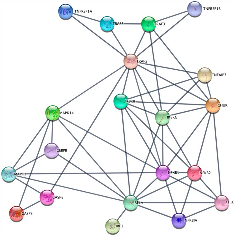 Fig.1 Reconstruction of the small gene network of the TNF-α signaling pathway from TNFR1 and TNFR2 in K562.1
The functional scope of TNFRSF1A crosses many physiological and pathological domains, echoing its ability to promote survival or destruction:
The complexities of TNFRSF1A, including ligand-dependent trimerization, PLAD-mediated pre-assembly, death domain–adaptor interactions, and sensitivity to metalloprotease shedding, complicate structural and pharmacological characterization. To address these challenges, Creative Biolabs has developed a custom TNFRSF1A protein design platform that provides receptor constructs suitable for structural biology, ligand-binding studies, and signaling research. Using structural insights and codon-optimized synthetic genes, our engineering team develops constructs tailored to specific experimental requirements while considering key structural and functional regions of TNFRSF1A. Every engagement begins with a thorough technical consultation to ensure that the protein architecture fits the intended application. Specific receptor formats, interaction interfaces, preparation strategies, and functional assays are determined according to individual project requirements.
Not finding the membrane protein product you need? Contact us to start your one-stop custom service!
Functional cellular models expressing TNFRSF1A on the cell surface can support agonist/antagonist screening, antibody evaluation, and studies of receptor-mediated signaling. Using optimized transduction and selection protocols, Creative Biolabs engineers custom TNFRSF1A stable cell lines with sustained receptor expression. The cell engineering platform utilizes lentiviral delivery, transposon-mediated integration, and targeted knock-in approaches depending on project requirements. Generated TNFRSF1A cell models may support studies of TNF responses, downstream signaling, and receptor shedding. Specific expression characteristics, phenotypic validation, and functional performance are documented according to the corresponding cell-line datasheet or individual project requirements.
Not finding the stable cell line product you need? Contact us to start your one-stop custom service!
To detect TNFRSF1A in inflamed tissue, delineate its surface distribution across cell types, selectively antagonize TNFR1 but not TNFR2 and neutralize the therapeutic effects of excess solubleTNF-αin vivo requires high-affinity sequence-defined antibodies to prevent receptor dimerization. We provides end-to-end recombinant antibody discovery programs focused on PLAD and CRD - Helix 2 regions of TNFRSF1A epitopes (extracellular & conformation directed). The pipeline includes immunogen design, multi-platform selection and downstream engineering to produce binders with the desired specificity affinity and developability profiles for research diagnostic or therapeutic applications. Using recombinant expression and clonal sequencing, we remove the batch-to-batch variability seen with conventional polyclonal sera to provide you a renewable fully characterized reagent.
Not finding the recombinant antibody product you need? Contact us to start your one-stop custom service!
Partnering with Creative Biolabs for TNFRSF1A protein engineering offers differential access to a vertically integrated platform underpinned by deep death receptor immunology expertise and best-in-class quality standards:
Yes, we provide recombinant proteins and stable cell lines with known mutations in the extracellular cysteine-rich domains that can be tested directly for comparison of ligand-binding affinity, receptor oligomerization or inflammatory signaling output to those from wildtype controls.
Yes, the protein is buffered at defined conditions without carrier proteins or primary amines in an appropriate buffer with immediate immobilization to sensor chips for TNF-α binding kinetics and therefore can be used directly after its purification without dialysis.
Yes, the Immunogen binds to a conserved region. This provides consistent detection across human, mouse and rat preparations without species-specific reagents.
Certainly, we have established two dual-stable lines constitutively expressing both full-length TNFRSF1A and TRADD where DISC formation & NF-κB activation kinetics can be assessed directly avoiding the variability associated with transient transfections.








