
Creative Biolabs is offering the most comprehensive services for antibody development projects. With strict regulation and effective execution, we are dedicated to providing the most valuable solutions to complete your projects.
HighlightsCreative Biolabs is offering the most comprehensive services for antibody development projects. With strict regulation and effective execution, we are dedicated to providing the most valuable solutions to complete your projects.
HighlightsCreative Biolabs has established a broad range of platforms for developing novel antibodies or equivalents. These cutting-edge technologies enable our scientists to meet your demands from different aspects and tailor the most appropriate solution that contributes to the success of your projects.
HighlightsWith deep understanding in antibody-related realms and extensive project experience, Creative Biolabs offers a variety of references to help you learn more about our capacities and achievements, including infographic, flyer, case study, peer-reviewed publications, and all kinds of knowledge that can assist your projects. You are also welcome to contact us directly for more specific solutions.
HighlightsGet a real taste of Creative Biolabs, one of the most professional custom service providers in the world. We are committed to providing highly customized comprehensive solutions with the best quality to advance your projects.
Highlights Loading...
All listed services and products are For Research Use Only. Do Not use in any diagnostic or therapeutic applications.
Tumor necrosis factor receptor superfamily 8(TNFRSF8) is a type I single transmembrane receptor encoded by the TNFRSF8 gene, which is mainly expressed on the surface of mature lymphocytes and myeloid cells in secondary immune tissues. The extracellular domain of the protein contains a number of highly conserved cysteine-rich ligand binding domains, with a single-segment transmembrane helix structure anda long intracellular cytoplasmic tail containing TRAF-binding regions, which can recruit TRAF-family adaptor proteins after ligand engagement and initiate downstream signal transduction programs. Structural studies show that the extracellular tandem cysteine repeats can form a specific ligand binding pocket, which can specifically recognize the homotrimeric secretory ligands of tumor necrosis factor superfamily. After the stable trimer assembly of the receptor, the intracellular signal cascade reaction can be started in order to mediate immune signal transmission.
In the absence of TNFRSF8 expression regulation, ligand-mediated signaling pathway will continue to be abnormally activated, leading to the disorder of basic activity of immune cells. Different lymphocyte subsets can differentially regulate the expression abundance of TNFRSF8 on the cell membrane surface, forming a gradient immune response threshold, effectively avoiding the persistent overactivation of immune cells under physiological homeostasis and maintaining the stability of immune microenvironment. There are gradient differences in ligand concentrations in various immune microenvironments, and the trimer assembly efficiency of TNFRSF8 can directly determine the conduction intensity of downstream intracellular signals. The extracellular cysteine repeating units can form independent stable folding structures through intramolecular disulfide bonds, which jointly determine the ligand recognition specificity of the receptor. In addition, the conservative cysteine pairing mode can stabilize the spatial conformation of the ligand binding pocket, ensure the reversible assembly and dynamic binding of the homotrimer ligand-receptor complex, and realize the accurate and controllable transmission of immune signals.
Variants within TNFRSF8 cysteine-rich regions could disrupt disulfide fold integrity and lower homotrimeric ligand binding affinity, which might alter lymphoid cell response thresholds in immune tissue model systems. No other TNF receptor paralog fully duplicates TNFRSF8’s combined ligand selectivity andcytoplasmic TRAF-mediated signaling capacity, though partial ligand overlap exists among superfamily members. TNFRSF8 localizes exclusively to outer plasma leaflets and only achieves full signal competence after ligand-induced homotrimer assembly. Its type I membrane topology separates extracellular ligand-sensing folds from cytoplasmic adaptor recruitment sequences, granting dual functional potential: extracellular cytokine detection and initiation of cytoplasmic immune signaling cascades. Reduced TNFRSF8 surface levels blunt ligand-triggered signal output and lower immune cell responsiveness to interstitial cytokine gradients, making this lymphoid receptor a suitable research target for immune threshold regulation analysis.
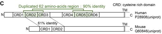 Fig. 1 Linear domain organization of human TNFRSF8, showing extracellular cysteine‑rich domains and transmembrane segment.1
The biological functions of integral membrane TNFRSF8 immune receptor are focused onhomotrimeric ligand capture and TRAF-family adaptor recruitment:
Creative Biolabs offers purified TNFRSF8 membrane protein samples produced under unified preparation workflows, including full-length TNFRSF8 constructs and isolated cysteine-rich extracellular variants. Truncated fragments cannot support complete ligand homotrimer assembly and adaptor recruitment activity, while full-length forms fit lymphoid immune research. All batches receive uniform quality screening. Functional assessments may only be performed under simulated lipid bilayer microenvironment setups. Cysteine repeat disulfide fold structural features are preserved across batches to support comparative ligand trimer binding analysis between experimental groups. Full-length TNFRSF8 membrane samples retain intact ligand coordination pockets post-purification, supporting reliable detection of transient receptor-ligand trimers in comparative functional analysis.
Not finding the Membrane Protein product you need? Contact us to start your one-stop custom service!
Creative Biolabs provides cell research models with adjustable TNFRSF8 expression levels, suitable for structural observation of type I TNF receptors and lymphoid cytokine research. Sample assessment covers sustained membrane detection and ligand trimer binding analysis, enabling side-by-side comparison of signal threshold behavior under varying TNFRSF8 abundances. These cell models can be paired with immune messenger quantification schemes to track response shifts linked to receptor dosage changes.
Not finding the stable cell line product you need? Contact us to start your one-stop custom service!
Anti-TNFRSF8 recombinant antibodies are generated via standardized workflows, compatible with lymphoid cell membrane localization mapping and ligand trimer complex identification. The antibody series supports multi-dimensional observation of TNFRSF8 distribution within secondary lymphoid tissue samples.
Not finding the recombinant antibody product you need? Contact us to start your one-stop custom service!
Beyond catalog products, Creative Biolabs offers specialized custom services for TNFRSF8 research:
TNFRSF8 may act as type I transmembrane TNF receptor to form homotrimers with matching secreted ligands and recruit cytoplasmic immune adaptor proteins.
TNFRSF8 surface density might adjust lymphoid cell cytokine responsiveness thresholds, serving as a key mediator of lymphoid tissue homeostasis biological processes.
No, all TNFRSF8 related products and services are strictly for research use only, and cannot be applied in clinical workflows. All material design and functional screening are optimized exclusively for basic laboratory research, without meeting clinical application criteria.
Offerings include full-length TNFRSF8 membrane protein, target-specific recombinant antibodies and tunable expression cell research models, supporting research on cytokine-triggered lymphoid signaling.
Laboratory analysis schemes may include homotrimeric cytokine co-binding assays to assess receptor cluster formation capacity under simulated lipid bilayer environments.








